The Health Education Assets Library (HEAL) is a collection of over 22,000 freely available digital materials for health sciences education. The collection is now housed at the University of Utah J. Willard Marriott Digital Library.
Filters: Collection: ehsl_heal
| Title | Description | Subject | Collection | ||
|---|---|---|---|---|---|
| 101 |
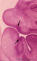 |
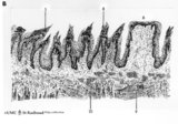
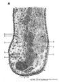
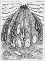
Sagittal section through tooth buds in jaws during tooth development - human, embryo, 6 weeks; low magnification | Stain: hematoxylin and eosin. At the top the upper jaw (maxilla) opposite the lower jaw (mandible). At the right side part of the tongue in the oral cavity. Arrows point to epithelial proliferations (future tooth bud formation) that are surrounded by condensation of mesenchymal cells (neural crest m... | oral cavity; tooth bud; tooth development | Poja Histology Collection - Oral Cavity Subset |
| 102 |
 |
Scheme of circumvallate papillae of the tongue (human, adult) | 1. (circum)vallate papilla; 2. non-keratinized stratified squamous epithelium; 3. taste bud; 4. serous gland (von Ebner); 5. draining duct (of serous gland); 6. lingual muscle | oral cavity; von Ebner; circumvallate papillae | Poja Histology Collection - Oral Cavity Subset |
| 103 |
 |
Scheme of compound glands in the oral cavity - human, adult | A. parotid gland (serous gland) magnification x 350 objective. B. sublingual gland (mucous part) magnification x 350 objective. C. sublingual gland (mixed part) magnification x 350 objective. D. simplified scheme of compound serous/mucous gland. 1. myoepithelial cell; 2. serous acinus; 3. stri... | oral cavity | Poja Histology Collection - Oral Cavity Subset |
| 104 |
 |
Scheme of cross sectional tooth in alveolar bone - cat | A. Survey of tooth (in alveolar bone) magnification x 30 objective. B. Peripheral part of tooth (in alveolar bone) magnification x 300 objective. 1. radicular pulp; 2. odontoblast layer; 3. dentin (radial arranged dentinal tubules); 4. Tomes' granular layer; 5. cementum; 6. fibers of period... | oral cavity; alveolar bone | Poja Histology Collection - Oral Cavity Subset |
| 105 |
 |
Scheme of filiform and fungiform papillae of the tongue - human, adult | 5. filiform papillae; 6. fungiform papillae; 8. locally cornification on top of filiform papillae; 9. lingual muscle; 10. lingual glands | oral cavity; papillae | Poja Histology Collection - Oral Cavity Subset |
| 106 |
 |
Scheme of tongue papillae and foliate papillae | A. Circumvallate papillae (tongue, human, adult) magnification x 35 objective. B. Foliate papillae (tongue, rabbit) magnification x 85 objective. C. Taste bud in foliate papilla (tongue, rabbit) magnification x 750 objective. 1. (circum)vallate papilla; 2. non-keratinized stratified squamous epit... | oral cavity; von Ebner; circumvallate papillae; foliate papillae | Poja Histology Collection - Oral Cavity Subset |
| 107 |
 |
Scheme of tongue papillae and taste buds | B. Scheme of foliate papillae of the tongue (rabbit) magnification x 85 objective. C. Scheme of taste bud in foliate papilla of the tongue (rabbit). 2. non-keratinized stratified squamous epithelium; 3. taste bud; 4. serous gland (von Ebner); 5. draining duct (of serous gland); 7. foliate papil... | oral cavity; von Ebner; foliate papillae | Poja Histology Collection - Oral Cavity Subset |
| 108 |
 |
Scheme of tooth development (human) | A. Survey of tooth germ (bell stage, embryo) magnification x 35 objective. B. Formation of enamel and dentin (bell stage, embryo) magnification x 350 objective. 1. pulp organ; 2. odontoblasts; 3. mantle predentin; 4. dentin; 5. enamel; 6. initial enamel; 7. inner dental epithelium (ameloblasts);... | oral cavity; dental lamina; bell stage | Poja Histology Collection - Oral Cavity Subset |
| 109 |
 |
Scheme survey dorsal surface of tongue - human, adult | 1. foliate papillae (rudimentary in human); 2. circumvallate papillae; 3. foramen cecum; 4. palatine tonsils; 5. filiform papillae; 6. fungiform papillae; 7. epiglottis; 11. lingual tonsils | oral cavity | Poja Histology Collection - Oral Cavity Subset |
| 110 |
 |
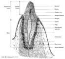
Scheme survey lip - human, adult | 1. red (transitional) zone; 2. keratinized stratified squamous epithelium (outer side); 3. non-keratinized stratified squamous epithelium (inner side); 4. hair shaft; 5. hair follicle; 6. sebaceous gland; 7. cross-section of circumoral muscle; 8. labial glands with draining ducts; ... | oral cavity | Poja Histology Collection - Oral Cavity Subset |
| 111 |
 |
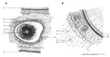
Scheme survey tooth (longitudinal section; human; canine) | Canine tooth in osseous alveolus. | oral cavity; pulp canal | Poja Histology Collection - Oral Cavity Subset |
| 112 |
 |
Scheme survey uvula - soft palate, human, adult | 1. nasal side - pseudostratified columnar ciliated epithelium (respiratory epithelium); 2. oral side - non-keratinized stratified squamous epithelium; 3. mixed glands (pharyngeal glands); 4. uvular muscle; 5. mucous palatine glands | oral cavity | Poja Histology Collection - Oral Cavity Subset |
| 113 |
 |
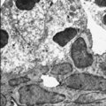
Secretory ameloblasts in tooth development - bell stage, gerbil, postnatal | Electronmicroscopy. Cross-sectioned distal sides of ameloblasts (secretion areas or Tomes' processes) with numerous organelles. The dark stained vesicles represent secretory granules contain among others amelogenin. At the right bottom corner intercellular spaces are filled with dark-stained organi... | oral cavity; enamel prisms | Poja Histology Collection - Oral Cavity Subset |
| 114 |
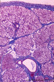
 |
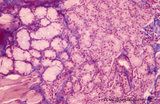
Sublingual gland (human) | Stain: Azan. The sublingual gland is a mixed gland and mucous cells are dominating (muco-serous acini). Intralobular ducts are present. Note one thick blue interlobular septum and few thinner intralobular ones. | oral cavity; seromucous gland | Poja Histology Collection - Oral Cavity Subset |
| 115 |
 |
Sublingual gland (human) | Stain: Azan. The sublingual gland is a mixed gland and characteristic in human is the presence of large areas of serous cells neighboring areas with mucous cells. Inter- and intra-lobular ducts are present. Note inter- and intra-lobular septa (blue) and scattered fat cells (white). | oral cavity; seromucous gland | Poja Histology Collection - Oral Cavity Subset |
| 116 |
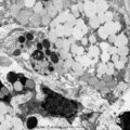 |
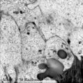
Submandibular gland (gerbil) | Electronmicroscopy. Upper two mucous cells of a seromucous acinus contain secretion droplets of different densities (light grey). Note partly fused mucous droplets. In the middle a serous cell cell with darkly stained granules. At the bottom a dense stained intercalated cell without granules. The ba... | oral cavity; seromucous gland | Poja Histology Collection - Oral Cavity Subset |
| 117 |
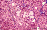 |
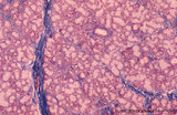
Submandibular gland (human) | Stain: Azan. The submandibular gland consists of mixed glands, i.e., the serous cells outnumber the mucous cells (seromucous acini). Locally crescent-shaped groups of serous cells (demi-lunes of Giannuzzi) are localized close to mucous cells to form an acinus.Intralobular ducts (left half) and thin ... | oral cavity; seromucous gland | Poja Histology Collection - Oral Cavity Subset |
| 118 |
 |
Submandibular gland (human) | Stain: Azan. Survey: on top capsule of dense connective tissue (blue) with septa dividing parenchyma of serous cells into lobules. At the bottom a large interlobular duct. Note white fat cells (adipocytes). | oral cavity; seromucous gland | Poja Histology Collection - Oral Cavity Subset |
| 119 |
 |
Submandibular gland (rat) | Electronmicroscopy. Darkly stained cells around the lumen of an intercalated duct and close to the lightly stained (partly degranulated) serous cells. | oral cavity; seromucous gland; intercalated duct | Poja Histology Collection - Oral Cavity Subset |
| 120 |
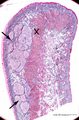 |
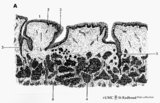
Survey of lip (human) | Stain: Azan. Arrows: labial glands in submucosa (mucous inner surface with non-keratinized epithelium). X: orbicularis oris muscle. Right side = skin surface (keratinized epithelium). Top = transitional zone (red zone) with slightly keratinized epithelium. | oral cavity; red zone; labial glands | Poja Histology Collection - Oral Cavity Subset |
| 121 |
 |
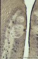
Taste bud of a foliate papil of the tongue (dorsal side, rabbit; high magnification) | Stain: Heidenhain's iron hematein. Taste bud with slender cells and darker stained spiky nuclei (type I cell), and cells with light stained round nuclei (type II cells). Below axons of the gustatory nerve fibers and nuclei of Schwann cells. Arrow points to narrow pore where dark stained fluffy struc... | oral cavity; foliate papillae; taste pore | Poja Histology Collection - Oral Cavity Subset |
| 122 |
 |
Taste bud of a foliate papilla of the tongue (dorsal side, human) | Stain: antikeratin-7 / immunoperoxidase and hematoxylin counterstained. Type I cells with small slender nuclei are identified by positive staining with cytokeratin 7 (monoclonal antibody OVTL12/30). | cytokeratin; oral cavity; foliate papilla; von Ebner | Poja Histology Collection - Oral Cavity Subset |
| 123 |
 |
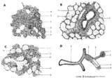
Taste bud of the tongue | Scheme electronmicroscopy. Generally a taste bud is composed of about 20 to 70 spindle-shaped epithelial cells. Slender type I cell ('Dark' cell) and oval shaped type II cell ('Light' cell) with a pale, round nucleus with their apices end in microvilli in the taste pore. The number of type I cells i... | oral cavity; foliate papillae; taste pore; electronmicroscopy | Poja Histology Collection - Oral Cavity Subset |
| 124 |
 |
Taste buds of foliate papillae of the tongue (dorsal side, rabbit) | Stain: Heidenhain's iron hematein. Taste buds within the lining (non-keratinized) epithelium of the groove. Note the pore in the left taste bud, with darkly stained fluffy apical microvilli. The lightly stained gustatory nerve fibers (nuclei of Schwann cells) approach the left taste buds. The taste ... | oral cavity; foliate papillae; taste pore | Poja Histology Collection - Oral Cavity Subset |
| 125 |
 |
Tongue (lingual gland, human) | Stain: Azan. The posterior lingual gland is composed of areas with pure serous acini neighbouring pure mucous acini between the tongue muscles (at the left few striated fibers). | oral cavity; lingual gland | Poja Histology Collection - Oral Cavity Subset |