Videos, clinical notes and related presentations concerning neuro-ophthalmological and neurovisual disorders collected during Dr. Wray's work as the Director of Neuro-Visual Disorders at Massachusetts General Hospital.
Shirley H. Wray, M.D., Ph.D., FRCP, Professor of Neurology Harvard Medical School, Director, Unit for Neurovisual Disorders, Massachusetts General Hospital.
NOVEL: https://novel.utah.edu/
TO
Filters: Collection: "ehsl_novel_shw"
| Title | History | Type | ||
|---|---|---|---|---|
| 101 |
 |
Latent Nystagmus | This is one of the first cases of latent nystagmus that I saw with Dr. Cogan in the 1970's. The presence of latent nystagmus was unknown to this patient, a little girl who was found to have latent nystagmus by the ophthalmologist at school trying to test the vision of each eye separately. When she... | Image/MovingImage |
| 102 |
 |
Lateropulsion | This 60 year old patient has Wallenberg's syndrome due to infarction of the left dorsolateral medulla. Wallenberg's syndrome is the best recognized syndrome involving the vestibular nuclei and adjacent structures. Unilateral infarcts affecting the vestibular nuclei may produce an oculomotor imbalanc... | Image/MovingImage |
| 103 |
 |
Lateropulsion | This 44 year old woman has a diagnosis of Multiple Sclerosis. | Image/MovingImage |
| 104 |
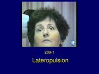 |
Lateropulsion | This 60 year old patient has Wallenberg's syndrome due to infarction of the left dorsolateral medulla. Wallenberg's syndrome is the best recognized syndrome involving the vestibular nuclei and adjacent structures. Unilateral infarcts affecting the vestibular nuclei may produce an oculomotor imbalanc... | Text |
| 105 |
 |
Lessons from the Bench and Bedside | Text | |
| 106 |
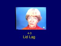 |
Lid Lag | The classical eye signs of thyroid associated ophthalmopathy (TAO) of Graves' Disease is illustrated by case ID925-4. This 50 year old woman with TAO is included in the collection because she illustrates very well lid lag (persistent elevation of the upper eyelid in downgaze) - von Graefe sign E... | Text |
| 107 |
 |
Lid Lag | The classical eye signs of thyroid associated ophthalmopathy (TAO) of Graves' Disease is illustrated by case ID925-4. This 50 year old woman with TAO is included in the collection because she illustrates very well lid lag (persistent elevation of the upper eyelid in downgaze) - von Graefe sign E... | Image/MovingImage |
| 108 |
 |
MS Time Lapse MRI | Professor Ian McDonald, Institute of Neurology, Queen Square, London contributed this remarkable Time-Lapse MRI of focal MS lesions in a single patient with multiple sclerosis over a period of one year. This time lapse video was assembled from serial T2- weighted MRI scans from a 25-year old wo... | Image/MovingImage |
| 109 |
 |
Midbrain Hemorrhage | The patient is a 49 year old woman who was in good health until January 17, 1991. When, at work one morning, she had an acute attack of light headedness and double vision and collapsed on the floor without loss of consciousness. She developed a severe retro-orbital headache. She was taken to t... | Text |
| 110 |
 |
Migraine / PET Study | In December 1994 the New England Journal of Medicine published a remarkable paper Bilateral Spreading Cerebral Hypoperfusion during Spontaneous Migraine Headache. Roger P. Woods, Marco Iacoboni and John C. Mazziotta. which is reproduced in part, and accompanied by a video illustration. Courtesy of J... | Image/MovingImage |
| 111 |
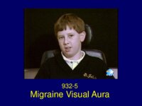 |
Migraine Visual Aura | The patient is a 9 year old right handed boy who developed headaches in 1993 at the age of 8. At that time he told his mother that he had bad headaches starting at the back of the head, usually bioccipital, spreading over the top of the head to his forehead. The headaches were short in duration la... | Text |
| 112 |
 |
Migraine Visual Aura | The patient is a 73 year old retired teacher who was referred in 1993 for a second opinion regarding treatment of episodic visual hallucinations. As a school boy in junior school, he began to experience transient episodes of a spot appearing in the right lower homonymous quadrant of his field of vi... | Image/MovingImage |
| 113 |
 |
Migraine Visual Aura | The patient is a 9 year old right handed boy who developed headaches in 1993 at the age of 8. At that time he told his mother that he had bad headaches starting at the back of the head, usually bioccipital, spreading over the top of the head to his forehead. The headaches were short in duration la... | Image/MovingImage |
| 114 |
 |
Migraine Visual Aura: A Discussion with Nobel Laureate David H. Hubel | I am greatly indebted to the Nobel Laureate, David Hubel for his permission to publish his description of his migraine aura. The recording was made fortuitously at the time that I invited David to the Unit for Neuro-Visual Disorders to record an audio clip describing the experiments in the cat that... | Image/MovingImage |
| 115 |
 |
Migraine Visual Aura: A Personal Account | I am greatly indebted to the Nobel Laureate, David Hubel for his permission to publish his description of his migraine aura. The recording was made fortuitously at the time that I invited David to the Unit for Neuro-Visual Disorders to record an audio clip describing the experiments in the cat that... | Image/MovingImage |
| 116 |
 |
Miller Fisher Syndrome (Guest Lecture) | Text | |
| 117 |
 |
Mitochondrial Myopathy (Guest Lecture) | Text | |
| 118 |
 |
Movement Disorders: A Brief Overview | Text | |
| 119 |
 |
Multiple Sclerosis | Text | |
| 120 |
 |
Multiple Sclerosis | The patient is a 56 year old woman who presented in 1982, at the age of 48, with a one week history of painless loss of vision in the left eye. Past History: Negative for a previous attack of optic neuritis or transient neurological symptoms. Family History: Negative for CNS disease Neuro-ophth... | Text |
| 121 |
 |
Multiple Sclerosis Lateropulsion | This 44 year old woman has a diagnosis of Multiple Sclerosis. | Text |
| 122 |
 |
Myasthenia Gravis (Guest Lecture) | The patient is a 46 year old woman who presented in July 1977 with horizontal double vision lasting two weeks. Three weeks later the left upper eyelid started to droop and by the end of the day the eye was closed. She had no ptosis of the right eye and no generalized fatigue. She consulted an in... | Text |
| 123 |
 |
Myasthenia Thymoma | The patient is a 46 year old woman who presented in July 1977 with horizontal double vision lasting two weeks. Three weeks later the left upper eyelid started to droop and by the end of the day the eye was closed. She had no ptosis of the right eye and no generalized fatigue. She consulted an in... | Text |
| 124 |
 |
Neonatal Opsoclonus | This child was one of the first cases of opsoclonus that I saw with Dr. Cogan in the early 1970's. The baby is a unique case in that in addition to neonatal opsoclonus with the characteristic multidirectional conjugate back-to-back saccades, periods of large amplitude upbeat nystagmus also occurred.... | Image/MovingImage |
| 125 |
 |
Neonatal Opsoclonus | This child was one of the first cases of opsoclonus that I saw with Dr. Cogan in the early 1970's. He carried a diagnosis of strabismus with deviation of the left eye. In this child, opsoclonus occurred as a transient phenomenon in an otherwise healthy infant. For a complete overview of opsoclonus i... | Image/MovingImage |
