John A. Moran Eye Center Neuro-Ophthalmology Collection: A variety of lectures, videos and images relating to topics in Neuro-Ophthalmology created by faculty at the Moran Eye Center, University of Utah, in Salt Lake City.
NOVEL: https://novel.utah.edu/
TO
Filters: Collection: "ehsl_novel_jmec"
| Title | Description | Type | ||
|---|---|---|---|---|
| 101 |
 |
Stereoacuity Testing | Demonstration of examination for stereoacuity. | Text |
| 102 |
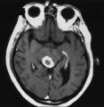 |
An Enhancing Bladder Metastasis Involving the Tectum of the Midbrain | Magnetic resonance image of an enhancing bladder metastasis involving the tectum of the midbrain of a 56-year-old man who developed double vision resulting from skew deviation and divergence insufficiency. He also had a left-sided relative afferent pupillary defect measuring 1.4 log units but showed... | Image |
| 103 |
 |
Ocular Myasthenia | Example of patient with myasthenia gravis. Demonstration of tensilon test. Patient shown to have bilateral ptosis, bilateral duction deficits, and left hypertropia. Discussion of techniques to observe subtle changes, such as bringing in a neutral observer or taking still photographs. Shows split-scr... | Image/MovingImage |
| 104 |
 |
Wall-Eyed Bilateral Internuclear Ophthalmoplegia (WEBINO) | Example of patient with horizontal binocular diplopia. Demonstration of exam, which shows alternating exotropia in cover test. As patient follows object, right eye does not pass the midline as the object moves to the left, while left eye go slightly past the midline, but does not abduct completely. ... | Image/MovingImage |
| 105 |
 |
Wall-Eyed Bilateral Internuclear Ophthalmoplegia (WEBINO) | Example of patient with Wall-Eyed Bilateral Internuclear Ophthalmoplegia. Patient is led through instructions for direction and distance of gaze. | Image/MovingImage |
| 106 |
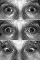 |
Light-near Dissociation | Light-near dissociation in a 51-year-old woman with multiple sclerosis who experienced double vision for 1 week. Her pupils are 5 mm in diameter in room light (top), react poorly in response to direct light reaction (middle), but constrict promptly in response to near stimulation (bottom). She also ... | Image |
| 107 |
 |
Migraine and Cluster Pathophysiology and Treatment | Video lecture covering pathophysiology and treatment of migraine and cluster headaches by Kathleen Digre, MD. | |
| 108 |
 |
The Normal Pupillary Light Reflex | The normal pupillary light reflex is initiated following exposure to light. After a brief latency, both the right (solid line) and left (broken line) pupil constrict, then undergo a small amount of redilation (escape), followed by oscillations (hippus) if the light is sustained. Hippus is not a path... | Image |
| 109 |
 |
Spontaneous Venous Pulsations | This clips shows a spontaneous venous pulsation viewed during an ocular examination. | Image/MovingImage |
| 110 |
 |
Duane's Retraction Syndrome Type 3 | Example of a patient with Type 3 Duane's Retraction Syndrome, as well as bilateral Duane's Syndrome. Shows limitation of abduction in both eyes and adduction in the left eye. Also shows side-view of globe retraction in abduction. | Image/MovingImage |
| 111 |
 |
Transillumination - Ciliary Body Neurofibromas | Example of transillumination on a patient with neurofibromatosis, but without Lisch nodules. Shows suspected neurofibromas in the ciliary body. | Image/MovingImage |
| 112 |
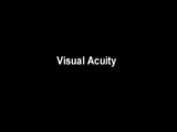 |
Measuring Visual Acuity | Demonstration on self of visual acuity exam, using a standard card. | Image/MovingImage |
| 113 |
 |
Spiral and Stellate Visual Fields Non-physiologic Variants | Description of testing the spiral and stellate visual fields. | |
| 114 |
 |
Introduction to Headache, Migraine and Secondary Headaches | Video lecture covering an introduction to headache, migraine, and secondary headaches by Kathleen Digre, MD. | |
| 115 |
 |
Transillumination - Lisch Nodules | Demonstration of transillumination of the Lisch nodules on a patient with neurofibromatosis. Shows how Lisch nodules that were not very visible in slit-lamp examination are better seen with transillumination, which may therefore be useful in detecting Lisch nodules earlier in children where they are... | Image/MovingImage |
| 116 |
 |
Introduction to the Basic Neurologic Exam | Introduction to the neurological examinations section of NExT. | |
| 117 |
 |
Nutritional Amblyopia | Example of patient with amblyopia with nutritional causes. | Text |
| 118 |
 |
Blepharospasm | Example of patient with blepharospasm. Patient is led through instructions for direction of gaze and opening and closing of eyes. Patient is led through same exercises again after receiving indomethacin treatment. | Image/MovingImage |
| 119 |
 |
4-54a -Optic Neuropathy, Ischemic: Posterior | Image | |
| 120 |
 |
Anterior Ischemic Optic Neuropathy | PPT describing Anterior Ischemic Optic Neuropathy (AION). Covers clinical signs, such as monocular vision loss, swollen nerve, and visual field defects, as well as risk factors. | Text |
| 121 |
 |
4-54b - Optic Neuropathy, Ischemic: Posterior | Image | |
| 122 |
 |
Optic Disc: Anatomy, Variants, Unusual discs | Discussion of viewing the optic disc. Includes development of direct ophthalmoscope. Covers normal optic disc and nerve fiber; nerve fiber loss and defects; cilioretinal arteries; venous anomolies; papilledema; pseudopapilledema; myopic disc; hyperoptic disc; little red discs; megallopapilla; myelin... | Text |
| 123 |
 |
3-3 - Bergmeister Papilla | Image | |
| 124 |
 |
3-4 - Tilted Disc | Tilted discs are normal variants caused by oblique insertion of the optic nerve to the globe. They can be and frequently are mistaken for papilledema. In this case the superior edge of the disc is tilted and appears elevated. This disc exhibits a nasal inferior tilt. | Image |
| 125 |
 |
3-5b - Myelinated Nerve Fibers | Myelinated nerve fibers are frequently confused with papilledema. The feathery edge of the myelinated fibers that conceal the disc and vessel should provide the clue. These myelinated nerve fibers make the disc look blurred. | Image |