The Health Education Assets Library (HEAL) is a collection of over 22,000 freely available digital materials for health sciences education. The collection is now housed at the University of Utah J. Willard Marriott Digital Library.
TO
Filters: Collection: "ehsl_heal"
| Title | Description | Subject | Collection | ||
|---|---|---|---|---|---|
| 101 |
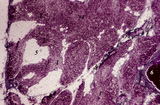 |
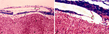
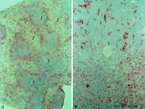
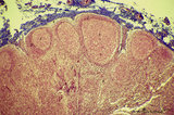
Age involution of thymus (human, postpuberal) | Stain: Azan. The size of the thymus is age-dependent, and undergoes a continuous process of involution, starting at postpuberal age. Due to depletion and reduced production of cortical thymocytes, as well as a gradual atrophy of the epithelial cells, the clear distinction between medulla (1) and cor... | thymus age; adipose cells; thymus involution; lymphoid tissue | Poja Histology Collection - Lymphatic Tissues and Organs Subset |
| 102 |
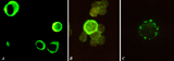 |
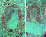
Immunohistochemical identification of splenic B cells (rat) | Stain: Immunofluorescence of FITC-labeled anti-Mark-1 antibody against rat B cells, carried out on spleen cells after fixation (A, B) or non-fixated spleen cells (C). (A) shows (young) plasma cells producing antibodies. (B) a green-stained B cell. The surrounding negative cells are likely T cells.... | immunofluorescence; B lymphocyte; capping | Poja Histology Collection - Lymphatic Tissues and Organs Subset |
| 103 |
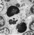 |
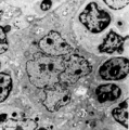
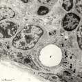
Thymus cortex (mouse, young adult) | Electron microscopy. Surrounded by thymocytes (2) a cortical macrophage (starry-sky macrophage) is seen and shows an electron-light nucleus (N) and a distinct nucleolus. The cell has engulfed two apoptotic thymocytes (1). The cytoplasm also contains small electron-dense lysosomes and myelin figures ... | cortical macrophage; epithelioreticular cell type II ; apoptotic thymocyte; lymphoid tissue | Poja Histology Collection - Lymphatic Tissues and Organs Subset |
| 104 |
 |
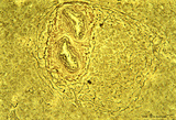
Survey of splenic trabecular artery (human) | Stain: Azan. The splenic artery (or lienal artery) is the blood vessel that provides oxygenated blood to the spleen. Branches of the splenic artery divide into trabecular arteries (1) which enter the white pulp as central arteries (4) that is surrounded with lymphocytes (5,periarteriolar lymphatic s... | trabecular artery; PALS; sinusoid | Poja Histology Collection - Lymphatic Tissues and Organs Subset |
| 105 |
 |
Immunohistochemistry of B cells in splenic white pulp (rat) | Stain: Immunohistochemistry of Vector red for Mark-1-antibody-stained B cells. (1) indicates the PALS area populated with unstained T cells located around the central artery of the white pulp. (2) indicates the positively red stained B cells in the germinal centre of the follicle. (3) the B-cells i... | marginal zone; immunohistochemistry; follicle | Poja Histology Collection - Lymphatic Tissues and Organs Subset |
| 106 |
 |
Penicillar arterioles in spleen (human) | Stain: Azan. The central or follicular artery in the follicle of the spleen splits into many arterioles. These arterioles spread as so-called penicillar arterioles (1) shown here. They are still surrounded by a very thin perilymphatic sheath (PALS) that disappears as the arteries in a brush-like pat... | penicillar arterioles; red pulp | Poja Histology Collection - Lymphatic Tissues and Organs Subset |
| 107 |
 |
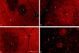
Localization of heparan sulfate (HS) in spleen (rat) | Stain: Immunofluorescence of Alexa 594 red labeled single chain antibody 3C3 for heparan sulfate (HS). The antibody stains HS epitopes of the meshwork of reticulum cells and the basal membrane of blood vessels. (1) central arteries in the T cell area of the PALS (2). The B cell area in the follic... | white pulp; heparan sulfate; PALS | Poja Histology Collection - Lymphatic Tissues and Organs Subset |
| 108 |
 |
Thymic nurse cell (TNC) (mouse) | Electron microscopy. The thymic nurse cell (TNC) consists of an epithelial reticular cell (type II) enclosing thymocytes. The TNC exists as a sealed structure in situ, i.e. the lymphocytes within the TNC are isolated from the general thymic environment. TNC are located in the cortex, where mature ... | epithelioreticular cell type II; thymic nurse cell | Poja Histology Collection - Lymphatic Tissues and Organs Subset |
| 109 |
 |
Scheme of lymph node (human) | Lymph node: A. survey; B. detail subcapsular (or marginal) sinus 1. adipose tissue; 2. capsule; 3. afferent lymph vessel with valves (B); 4. subcapsular (or marginal) sinus; 5. germinal centre of a secondary lymphatic nodule (or follicle); 6. crescent or mantle zone indicated by cap of per... | cortex; scheme; paracortex; germinal center | Poja Histology Collection - Lymphatic Tissues and Organs Subset |
| 110 |
 |
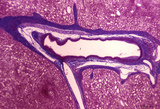
Afferent lymph vessel in lymph node (human) | Stain: Azan. Left (A) and right (B): part of the cortex of a lymph node with the capsule (1) and subcapsular (or marginal) sinus (2) filled with lymphocytes. Left (A): (3) show perpendicularly localized reticular cells and fibres (blue) in the sinus. (4) indicate afferent lymph vessel with valves ... | lymph vessel; subcapsular sinus; follicle; cortex | Poja Histology Collection - Lymphatic Tissues and Organs Subset |
| 111 |
 |
The effect of cyclophosphamide on splenic B cells (rat) | Stain: Immunoperoxidase staining using diaminobenzidin (DAB)/ hematoxylin counterstained on frozen section of B cells with the antibody Mark-1. The PALS area (1) contains T cells and remains unstained blue. The positively stained B cells (brown) are found in the germinal centres (2) and in the coron... | cyclophosphamide; immunosupression; B lymphocytes; Mark 1 antibody | Poja Histology Collection - Lymphatic Tissues and Organs Subset |
| 112 |
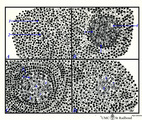 |
Scheme of development of germinal centre in lymphatic nodule (human) | A. primary nodule (or follicle) with mainly nave B lymphocytes and some memory cells, but no germinal centre; B. proliferation of lymphoblasts by mitosis (3,-->) and development of capillaries (*) in the centre of the follicle; C. so-called secondary nodule with lighter stained germinal centre wit... | follicle; antigen stimulation; scheme; germinal center | Poja Histology Collection - Lymphatic Tissues and Organs Subset |
| 113 |
 |
Age involution of thymus (human) | Stain: Azan. A: Although the adipose tissue in the thymus of a patient of 65 years is predominant it still contains areas of remnants (arrows 1+2) of cortical (2) and medullary portions (1) of the thymus. B+C: Degradation of thymic tissues is less progressed in adults and shows less replacement of... | thymus age; thymus involution; adipose cells; lymphoid tissue | Poja Histology Collection - Lymphatic Tissues and Organs Subset |
| 114 |
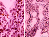 |
Splenic sinusoids (human) | Stain: A: Azan and B: Silver stain (Gomori). The splenic sinusoids are built in the form of a grid. The frame of the barrel consists of elongated endothelial cells (1) which are spirally wrapped around by reticular fibers (2). To this reticular fibers are attached many macrophages which control the... | sinusoid; reticular fibers | Poja Histology Collection - Lymphatic Tissues and Organs Subset |
| 115 |
 |
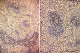
Circumscript reticular pattern of splenic lymphatic nodule (human) | Stain: Silver stain (Gomori). In this particular case the black-stained reticular network is beautiful organized in and around the nodule. Note also the reticulin staining between the myocytes in the wall of both cross-sectioned (1) central arteries. (2) follicle with lymphocytes. | sinusoid; reticular fibers; follicle | Poja Histology Collection - Lymphatic Tissues and Organs Subset |
| 116 |
 |
High endothelial venules (HEV) (rat lymph node, human palatine tonsil) | Stain: (A) Anti-laminin-antibody and immunoperoxidase staining using diaminobenzidin (DAB)/ hematoxylin counterstained on frozen section of lymph node; (B) electron microscopy (palatine tonsil). (A): Using an antibody against laminin brown-stained basement membranes are outlined demonstrating postc... | palatine tonsil; high endothelial venule (HEV); laminin; immunohistochemistry | Poja Histology Collection - Lymphatic Tissues and Organs Subset |
| 117 |
 |
Thymus cortex (rat, young adult) | Electron microscopy. Type I epithelioreticular cells separate connective tissue compartment from the thymic parenchyma. With occludens junctions and desmosomes as barriers they form wide-mesh networks creating specific microenvironments for developing T cells. The extensions of type I cells (5) are... | thymus cortex; epithelioreticular cell type I; epithelioreticular cell type II; lymphoid tissue | Poja Histology Collection - Lymphatic Tissues and Organs Subset |
| 118 |
 |
Immunohistochemistry with cellular markers in thymus (rat) | Stain: Alexa 594 red immunofluorescence. (1) medulla; (2) cortex; (3) septa. (A): Strong CD8 staining of the thymic cortical lymphocytes (single CD8+ and double positive CD4+CD8+ thymocytes). (B): OX19 staining for almost all T cells, equivalent to T1 human marker and Ly-1 mouse marker. Note that... | cyclophosphamide; CD8 monoclonal antibody; OX19 monoclonal antibody; ER1 monoclonal antibody | Poja Histology Collection - Lymphatic Tissues and Organs Subset |
| 119 |
 |
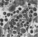
Hassall's corpuscle in thymus (mouse) | Electron microscopy. The centre of a small Hassall's corpuscle consists of darker-stained cells (1) which are keratinizing and represent degenerating cytoplasmic remnants (2). (3) is an infiltrating monocyte and (*) indicate the presence of free keratohyalin granules from disintegrated epithelial ce... | thymic corpuscle (Hassalls); lymphoid tissue ; epithelioreticular cell (ERC) | Poja Histology Collection - Lymphatic Tissues and Organs Subset |
| 120 |
 |
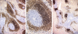
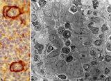
MHC-class II dendritic cells in spleen (rat) | Stain: Immunohistochemistry of Vector red staining of MHC-class II with ER13 antibody. The survey in (A) shows that MHC-class II expressing cells are conspicuously concentrated in dendritic cell types (B) in the transition zone between red pulp (2) and white pulp (1). These dendritic or antigen-pres... | MHC class II; dendritic cells; immunofluorescence | Poja Histology Collection - Lymphatic Tissues and Organs Subset |
| 121 |
 |
Thymus medulla (rat, young adult) | Electron microscopy. Epithelioreticular cell of the medulla with an electron-light cytoplasm contains cross-sections of vacuoles with small finger-like cytoplasmic extrusions in the lumen (1). Electron-dense lysosomal structures (2) are also present as well as bundles of intermediate filaments (kera... | medullar epithelioreticular cell ; keratin filaments; lymphoid tissue | Poja Histology Collection - Lymphatic Tissues and Organs Subset |
| 122 |
 |
Immunoperoxidase stained CD3 positive T cells in spleen (rat) | Stain: Immunoperoxidase staining using diaminobenzidin (DAB)/ hematoxylin counterstained on frozen section with anti CD3 antibodies. Survey (A) and detail (B) show that CD3 marker for mature T cells stains positively (brown) in the PALS area (1) around the central arteries, while the adjacent follic... | CD3 lymphocytes; immunohistochemistry; PALS | Poja Histology Collection - Lymphatic Tissues and Organs Subset |
| 123 |
 |
: Lymph node (rat) | Electron microscopy. A low magnification of a part of the medulla showing medullary cords surrounded by labyrinthine medullary sinus (*). In this picture the medullar cord runs from left bottom corner to right top corner, and is lined by flat reticular cell types. Within the cord one finds a star-sh... | medulla; electron microscopy; sinusoid | Poja Histology Collection - Lymphatic Tissues and Organs Subset |
| 124 |
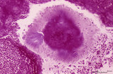 |
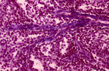
Calcified bacterial plaque in palatine tonsil ('gut-associated lymphatic tissue' or GALT) (human) | Stain: Azan. A tonsillar crypt lined by squamous epithelium (2) that is infiltrated with lymphocytes (1). It is a normal finding that within the crypts free cells, plugs of lymphocytes (1) and calcified epithelial debris as well as colonies of oral commensally bacteria are present. (3) shows a calc... | bacterial plaque | Poja Histology Collection - Lymphatic Tissues and Organs Subset |
| 125 |
 |
Cortex of lymph node (human) | Stain: Azan. The cortex of the lymph node comprises the (secondary) follicles with germinal centres (1) and corona or mantle zone (2), all filled with B lymphocytes. The paracortical area (3) below the follicles largely comprises T cells. Afferent lymph vessels enter via the capsule (6) into the m... | cortex; paracortex | Poja Histology Collection - Lymphatic Tissues and Organs Subset |