The Health Education Assets Library (HEAL) is a collection of over 22,000 freely available digital materials for health sciences education. The collection is now housed at the University of Utah J. Willard Marriott Digital Library.
TO
Filters: Collection: "ehsl_heal"
| Title | Description | Subject | Collection | ||
|---|---|---|---|---|---|
| 101 |
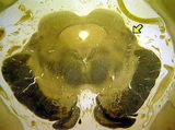 |
Brachium of inferior colliculus | Brachium of inferior colliculus. Midbrain. Transverse plane. Photograph. Multimedia. | Inferior colliculus; Mesencephalon; Central nervous system; Anatomy | Slice of Life |
| 102 |
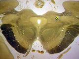 |
Brachium of superior colliculus | Brachium of superior colliculus. Pretectum-superior colliculus. Transverse plane. Photograph. Multimedia. | Superior colliculus; Mesencephalon; Tegmentum mesencephali; Central nervous system; Anatomy | Slice of Life |
| 103 |
 |
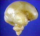
Brain 32 weeks, normal, lateral view, lateral fissure, insula | Brain 32 weeks, normal, lateral view, lateral fissure, insula. Lateral view. Photograph. Multimedia. | Brain; Fetus; Central nervous system; Anatomy | Slice of Life |
| 104 |
 |
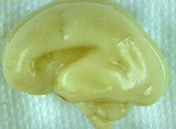
Brain 32 weeks, normal, midsagittal | Brain 32 weeks, normal, midsagittal. Sagittal plane. Photograph. Multimedia. | Brain; Fetus; Central nervous system; Anatomy | Slice of Life |
| 105 |
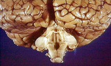 |
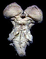
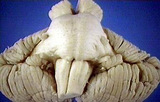
Brain stem posterior view, closeup fourth ventricle, cerebellum removed showing all cerebellar peduncles | Brain stem posterior view, closeup fourth ventricle, cerebellum removed showing all cerebellar peduncles | Central Nervous System; Brain Stem; Cerebellum | Slice of Life |
| 106 |
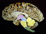 |
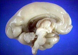
Brain stem, all regions midsagittal section | Brain stem, all regions midsagittal section. With cerebellum too. Graphic overlay. Sagittal plane. Photograph. Multimedia. | Brain stem; Cerebellum; Brain; Central Nervous System; Anatomy | Slice of Life |
| 107 |
 |
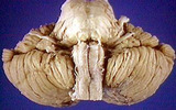
Brain stem, dorsal surface | Brain stem, dorsal surface. Used for graphic overlay (digitized image). Photograph. Multimedia. | Brain stem; Brain; Central nervous system; Anatomy | Slice of Life |
| 108 |
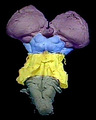 |
Brain stem, dorsal surface, each region | Brain stem, dorsal surface, each region. All regions colored, graphic overlay. Photograph. Multimedia. | Brain stem; Brain; Central nervous system; Anatomy | Slice of Life |
| 109 |
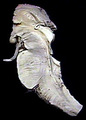 |
Brain stem, midsagittal | Brain stem, midsagittal. Used for graphics overlay. Sagittal plane. Photograph. Multimedia. | Brain stem; Brain; Central nervous system; Anatomy | Slice of Life |
| 110 |
 |
Brain stem, midsagittal regions | Brain stem, midsagittal regions. Graphics overlay. Sagittal plane. Photograph. Multimedia. | Brain stem; Central nervous system; Anatomy | Slice of Life |
| 111 |
 |
Brain stem, ventral surface | Brain stem, ventral surface. Photograph. Multimedia. | Brain Stem; Brain; Central Nervous System; Anatomy | Slice of Life |
| 112 |
 |
Brain stem, ventral surface | Brain stem, ventral surface. Without meninges. Photograph. Multimedia. | Brain stem; Meninges; Brain; Central nervous system; Anatomy | Slice of Life |
| 113 |
 |
Brain stem, ventral surface, unfixed | Brain stem, ventral surface, unfixed. Ventral surface. Photograph. Multimedia. | Brain stem; Brain; Central nervous system; Anatomy | Slice of Life |
| 114 |
 |
Brain, 15 1/2 week, normal human fetus | Brain, 15 1/2 week, normal human fetus. Ventral view. Photograph. Multimedia. | Brain; Fetus; Central nervous system; Anatomy | Slice of Life |
| 115 |
 |
Brain, 15 1/2 week, normal human fetus | Brain, 15 1/2 week, normal human fetus. Lateral view. Photograph. Multimedia. | Brain; Fetus; Central nervous system; Anatomy | Slice of Life |
| 116 |
 |
Brain, 22 week fetus | Brain, 22 week fetus. Normal, lateral view. Photograph. Multimedia. | Brain; Fetal development; Central nervous system; Anatomy | Slice of Life |
| 117 |
 |
Brain, 23 week, normal | Brain, 23 week, normal. Midsagittal view, choroid plexus can be seen in lateral and third ventricle. Sagittal plane. Photograph. Multimedia. | Brain; Choroid plexus; Fetus; Central nervous system; Cerebral ventricles; Anatomy | Slice of Life |
| 118 |
 |
Brain, 24 weeks, normal | Brain, 24 weeks, normal. Ventral view. Photograph. Multimedia. | Brain; Fetus; Central Nervous System; Anatomy | Slice of Life |
| 119 |
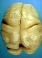 |
Brain, 24 weeks, normal | Brain, 24 weeks, normal. Dorsal view. Photograph. Multimedia. | Brain; Fetus; Central Nervous System; Anatomy | Slice of Life |
| 120 |
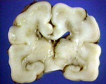 |
Brain, 27 week, normal | Brain, 27 week, normal. Coronal view with germinal plate and hippocampus. Coronal plane. Photograph. Multimedia. | Brain; Hippocampus; Fetus; Central nervous system; Anatomy | Slice of Life |
| 121 |
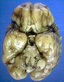 |
Brain, 27 week, normal human | Brain, 27 week, normal human. Ventral view. Photograph. Multimedia. | Brain; Fetus; Central nervous system; Anatomy | Slice of Life |
| 122 |
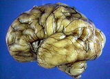 |
Brain, 27 week, normal human | Brain, 27 week, normal human. Lateral view of developing middle cerebral artery. Photograph. Multimedia. | Brain; Cerebral arteries; Central nervous system; Fetus; Anatomy | Slice of Life |
| 123 |
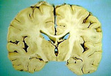 |
Brain, coronal thalamus and cortex, classical section | Brain, coronal thalamus and cortex, classical section. Coronal plane. Photograph. Multimedia. | Brain; Thalamus; Cerebral cortex; Central nervous system; Anatomy | Slice of Life |
| 124 |
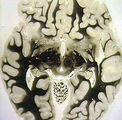 |
Brain, horizontal section | Brain, horizontal section. Level of superior colliculus. Horizontal plane. Photograph. Multimedia. | Brain; Superior colliculus; Central nervous system; Anatomy | Slice of Life |
| 125 |
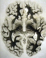 |
Brain, horizontal section | Brain, horizontal section. Level posterior commissure. Horizontal plane. Photograph. Multimedia. | Brain; Central nervous system; Anatomy | Slice of Life |