Home
Browse
Ask Us
Chat
Harmful Language Statement
Log in
Advanced Search
Year
1906
1907
1908
1909
1910
1911
1912
1913
1914
1915
1916
1917
1918
1919
1920
1921
1922
1923
1924
1925
1926
1927
1928
1929
1930
1931
1932
1933
1934
1935
1936
1937
1938
1939
1940
1941
1942
1943
1944
1945
1946
1947
1948
1949
1950
1951
1952
1953
1954
1955
1956
1957
1958
1959
1960
1961
1962
1963
1964
1965
1966
1967
1968
1969
1970
1971
1972
1973
1974
1975
1976
1977
1978
1979
1980
1981
1982
1983
1984
1985
1986
1987
1988
1989
1990
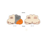
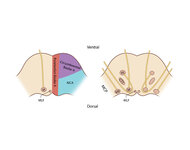
1991
1992
1993
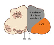
1994
1995
1996
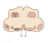
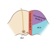
1997
1998
1999
2000
2001
2002
2003
2004
2005
2006
2007
2008
2009
2010
2011
2012
2013
2014
2015
2016
2017
2018
2019
2020
2021
2022
2023
2024
2025
TO
1906
1907
1908
1909
1910
1911
1912
1913
1914
1915
1916
1917
1918
1919
1920
1921
1922
1923
1924
1925
1926
1927
1928
1929
1930
1931
1932
1933
1934
1935
1936
1937
1938
1939
1940
1941
1942
1943
1944
1945
1946
1947
1948
1949
1950
1951
1952
1953
1954
1955
1956
1957
1958
1959
1960
1961
1962
1963
1964
1965
1966
1967
1968
1969
1970
1971
1972
1973
1974
1975
1976
1977
1978
1979
1980
1981
1982
1983
1984
1985
1986
1987
1988
1989
1990
1991
1992
1993
1994
1995
1996
1997
1998
1999
2000
2001
2002
2003
2004
2005
2006
2007
2008
2009
2010
2011
2012
2013
2014
2015
2016
2017
2018
2019
2020
2021
2022
2023
2024
2025
Type
Image/MovingImage
349
Image
67
Text
60
Image/StillImage
1
Format
video/mp4
347
application/pdf
68
image/jpeg
63
Institution
Eccles Health Sciences Library
463
University of Utah Partnerships
11
Department of Cultural and Community ...
1
Utah American Indian Digital Archive
1
Utah Museum of Natural History
1
University of Utah Marriott Library
1
More
Collection
Angus Munn Woodbury Papers
1
College of Architecture + Planning
3
College of Law Publications
6
NOVEL
3
NOVEL - Daniel Gold Collection
373
NOVEL - Daniel Gold Textbook
46
NOVEL - Frank B. Walsh Collection
7
NOVEL - Journal of Neuro-Ophthalmology
10
NOVEL - NANOS Annual Meeting
24
Peoples of Utah Revisited
1
UAIDA Main Collection
1
Undergrad Research Abstracts Journal
2
Vertebrate Zoology Field Notes
1
More
76
-
100
of
478
<
1
2
3
4
5
6
7
8
9
10
>
Gallery view
Number of results to display per page
10
25
50
100
200
Sort by Relevance
Sort by Title A-Z
Sort by Title Z-A
Sort by Date Ascending
Sort by Date Descending
Sort by Last Modified Ascending
Sort by Last Modified Descending
Title
Date
Type
Setname
76
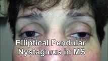
Elliptical Pendular Nystagmus in MS
2016
Image/MovingImage
ehsl_novel_gold
77
Enhanced Ptosis in Myasthenia Gravis
2016
Image/MovingImage
ehsl_novel_gold
78
The Episodic Vestibular Syndrome
2017
Image/MovingImage
ehsl_novel_gold
79
Examples of Patients with Saccadic Intrusions (Square Wave Jerks)
2017
Image/MovingImage
ehsl_novel_gold
80
Expanded Acute Onset Persistent Vision Loss Differential
Text
ehsl_novel_gold
81
Expanded Nystagmus & Saccadic Intrusions/Oscillations Differential
2021
Text
ehsl_novel_gold
82
Eye Closure and Oculopalatal Tremor
2017
Image/MovingImage
ehsl_novel_gold
83
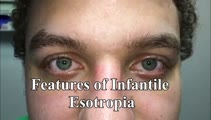
Eye Signs in Infantile Esotropia - Latent Nystagmus and Inferior Oblique Overaction
Image/MovingImage
ehsl_novel_gold
84
Eyelid Anatomy
2017
Image
ehsl_novel_gold
85
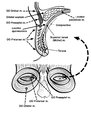
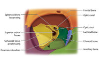
Figure 17: Bony Structures Relevant to the Orbit
2022
Image
ehsl_novel_gold
86
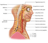
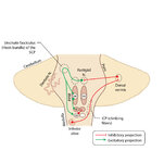
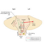
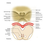
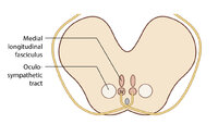
Figure 1: Oculosympathetic Pathway for Pupillary Dilation
2022
Image
ehsl_novel_gold
87
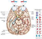
Figure 24: Typical Visual Field Defects Associated with Discrete Lesions Along the Visual Pathways
2022
Image
ehsl_novel_gold
88
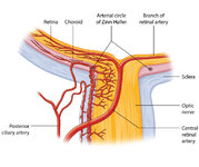
Figure 27: Vascular Supply of the Optic Nerve Head, Choroid and Retina
2022
Image
ehsl_novel_gold
89
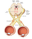
Figure 2: Parasympathetic Pathway for Pupillary Constriction
2022
Image
ehsl_novel_gold
90
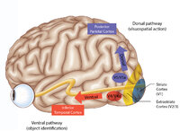
Figure 43: How the Brain Makes Sense of What It Sees - The Dorsal and Ventral Visual Pathways, and a 3 Tiered Approach to Vision
2022
Image
ehsl_novel_gold
91
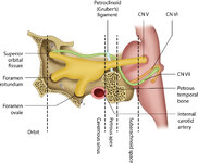
Figure 46: The Course of the 6th (VI) Nerve
2022
Image
ehsl_novel_gold
92
Figure 50: Anatomy and Physiology of the Saccadic Pathways
2022
Image
ehsl_novel_gold
93
Figure 51: Lateral Medullary Lesion Causing Saccadic Dysmetria
2022
Image
ehsl_novel_gold
94
Figure 51: Lateral Medullary Lesion Causing Saccadic Dysmetria (Supplement)
Image
ehsl_novel_gold
95
Figure 51: Lateral Medullary Lesion Causing Saccadic Dysmetria (Supplement)
Image
ehsl_novel_gold
96
Figure 53: Vascular Distribution and Anatomy Relevant to the Lateral Medullary (Wallenberg) Syndrome
2022
Image
ehsl_novel_gold
97
Figure 53: Vascular Distribution and Anatomy Relevant to the Lateral Medullary (Wallenberg) Syndrome (Supplement)
Image
ehsl_novel_gold
98
Figure 53: Vascular Distribution and Anatomy Relevant to the Lateral Medullary (Wallenberg) Syndrome (Supplement)
Image
ehsl_novel_gold
99
Figure 61: Vascular Distribution and Anatomy (Including 6th, 7th, 8th Nerves, MLF) of the Pons
2022
Image
ehsl_novel_gold
100
Figure 61: Vascular Distribution and Anatomy (Including 6th, 7th, 8th Nerves, MLF) of the Pons (Supplement)
Image
ehsl_novel_gold
76
-
100
of
478
<
1
2
3
4
5
6
7
8
9
10
>