The Health Education Assets Library (HEAL) is a collection of over 22,000 freely available digital materials for health sciences education. The collection is now housed at the University of Utah J. Willard Marriott Digital Library.
TO
Filters: Collection: ehsl_heal
| Title | Description | Subject | Collection | ||
|---|---|---|---|---|---|
| 76 |
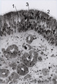 |
Olfactory mucosa in the nose (human) | Stain: Toluidine blue, one-micron Epon plastic section. Pseudostratified epithelium with light stained nuclei (1) (note distinct nucleoli) of olfactory cells. At the surface faintly stained cilia/microvilli (2). Nuclei of sustentacular (supporting) cells (3) are stained darker and predominantly in t... | Bowman glands; Olfactory glands; Olfactory mucosa; Pseudostratified epithelium | Poja Histology Collection - Respiratory System Subset |
| 77 |
 |
Olfactory vesicle (bulb) in the nose (gerbil) | DUPLICATE RECORD - FOR DELETION Electron microscopy. A distinct olfactory bulb (1, vesicle) with horizontally extended cross-sectioned cilia (*) and basal bodies (↑) is surrounded by numerous microvilli and free olfactory cilia. Close to the first bulb a second one is about to protrude above th... | Olfactory epithelium; Olfactory vesicle | Poja Histology Collection - Respiratory System Subset |
| 78 |
|
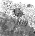
Olfactory vesicle (bulb) in the nose (gerbil) | Electron microscopy. A distinct olfactory bulb (1, vesicle) with horizontally extended cross-sectioned cilia (*) and basal bodies (↑) is surrounded by numerous microvilli and free olfactory cilia. Close to the first bulb a second one is about to protrude above the surface of the supporting cells b... | Olfactory epithelium; Olfactory vesicle | Poja Histology Collection - Respiratory System Subset |
| 79 |
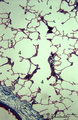 |
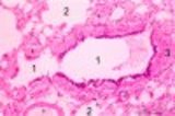
Peripheral alveolar area of the lung (human) | Stain: Azan. (1) Part of a pulmonary artery in a septum (2). (3) represents part of a bronchiolus respiratorius that continues into several alveolar ducts (4) and subsequently in alveolar sacs. Arrows (↓) indicate small foci of carbon deposits. | Respiratory bronchioli; Alveolar ducts; Alveolar sacs | Poja Histology Collection - Respiratory System Subset |
| 80 |
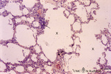 |
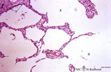
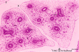
Progressing centrilobular lung emphysema (human, adult) | Stain: Hematoxylin and eosin. Emphysema is defined as enlargement of the air spaces (X) distal to the terminal bronchioles, with destruction of the alveolar walls. The remaining alveolar walls are thickened (1). There is an increased cellularity and locally signs of chronic inflammation (↓) are pr... | Poja Histology Collection - Respiratory System Subset | |
| 81 |
 |
Progressing centrilobular lung emphysema (human, adult) | Stain: Hematoxylin and eosin. Emphysema is defined as enlargement of the airspaces (X) distal to the terminal bronchioles, with destruction of the alveolar walls. At (3) remnant of a respiratory bronchiolus. Alveolar walls are destroyed and other alveolar walls are thickened (1) and show an increas... | Alveolar tips | Poja Histology Collection - Respiratory System Subset |
| 82 |
 |
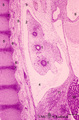
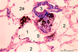
Pseudoglandular - canalicular period of developing lung (human, fetus) | Stain: Azan. Part of a bronchus (1) with young cartilage (2). The hyaline cartilage presents solitary chondrocytes embedded in the blue intercellular substance surrounded by a perichondrium (3). Note that the bronchus as well as the bronchial tubes (4) show an apical position of the epithelial nucle... | Lung development; Pseudoglandular period; Canalicular period; Mesenchyme | Poja Histology Collection - Respiratory System Subset |
| 83 |
 |
Pseudoglandular - canalicular period of developing lung (human, fetus) | Stain: Azan. Terminal budding and elongation of the future bronchial tree: branching of a bronchial tubes (1) within an islet where the mesenchyme condenses (4). A large blood vessel (2) as well as a lymph vessel (3) are recognizable. Note in the mesenchyme formation of blood capillaries in the vici... | Lung development; Pseudoglandular period ; Canalicular period; Mesenchyme | Poja Histology Collection - Respiratory System Subset |
| 84 |
 |
Pseudoglandular - canalicular period of developing lung (human, fetus) | Stain: Azan. Location of a cartilagineous ring (1) between two cross-sectioned bronchi (2). Note in the bronchial epithelial cells the apical position of the epithelial nuclei and the light-stained basal part (↑) containing glycogen. The young hyaline cartilage presents solitary chondrocytes embed... | Lung development; Pseudoglandular period; Canalicular period; Mesenchyme | Poja Histology Collection - Respiratory System Subset |
| 85 |
 |
Pseudoglandular - canalicular period of developing lung (human, fetus) | Stain: Azan. Longitudinal section through a large bronchus (1) with cartilagineous rings (2). At (3) developing glandular structures in islets of bronchial tubes surrounded by condensed mesenchyme. At (4) lymph vessels. | Lung development; Pseudoglandular period; Canalicular period; Mesenchyme; Bronchial tubes | Poja Histology Collection - Respiratory System Subset |
| 86 |
 |
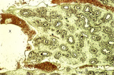
Pseudoglandular - canalicular period of developing lung (human, fetus) | Stain: Azan. Part of a future lung lobe with cross-sectioned larger bronchi (1) in close association with islets of future smaller bronchial tubes (*). Note the branching of the bronchial tubes within an islet. The mesenchyme within the islets of future tubes becomes more condensed (↓). Blood vess... | Lung development; Visceral pleura; Pseudoglandular period; Canalicular period; Mesenchyme | Poja Histology Collection - Respiratory System Subset |
| 87 |
 |
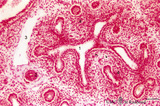
Pseudoglandular period of developing lung (human, embryo) | Stain: Trichrome (Goldner). Cross-sectioned future bronchial tubes (1) of varying sizes, note the apical position of the nuclei with light-stained basal parts (glycogen). The surrounding mesenchyme condenses (↓) around the epithelium and in the neighbourhood small blood vessels (*) are present. At... | Lung development; Pseudoglandular period; Bronchial tubes; Mesenchyme | Poja Histology Collection - Respiratory System Subset |
| 88 |
 |
Pseudoglandular period of developing lung (human, embryo) | Stain: Trichrome (Goldner). Cross-sectioned future bronchial tubes (1) of varying sizes, note the apical position of the nuclei with light-stained basal parts (glycogen). The surrounding mesenchyme becomes condensed (↓) around the epithelium and in between small blood vessels (*) are present. | Lung development; Pseudoglandular period; Bronchial tubes; Mesenchyme | Poja Histology Collection - Respiratory System Subset |
| 89 |
 |
Pseudoglandular period of developing lung (human, embryo) | Stain: Hematoxylin and eosin. Two cross-sectioned future bronchial tubes. Note the basal position of the nuclei with light-stained apical cytoplasm (↓, glycogen). The surrounding mesenchyme condenses (1) around the epithelium and developing capillaries and small blood vessels (*) are present. Futu... | Lung development; Visceral pleura; Pseudoglandular period; Bronchial tubes; Mesenchyme | Poja Histology Collection - Respiratory System Subset |
| 90 |
 |
Pseudoglandular period of developing lung (human, embryo, low magnification) | Stain: Hematoxylin and eosin. The two future lung lobes contain many cross-sectioned future bronchial tubes (1). Note in these epithelial cells the apical position of the nuclei with light-stained basal parts (glycogen). The surrounding mesenchyme becomes more condensed (↓) around the epithelium a... | Lung development; Visceral pleura; Pseudoglandular period; Bronchial tubes; Mesenchyme | Poja Histology Collection - Respiratory System Subset |
| 91 |
 |
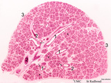
Pseudoglandular period of developing lung (human, embryo, low magnification) | Stain: Hematoxylin and eosin. Cross-sectioned future bronchial tubes (1), the surrounding mesenchyme becomes more condensed around the epithelium. The mesoderm of the future visceral pleura (2) as well as the future parietal pleura (3) and (4) indicates pleural cavity. The cartilagineous spinal colu... | Lung development; Visceral pleura; Parietal pleura; Pseudoglandular period; Bronchial tubes; Mesenchyme | Poja Histology Collection - Respiratory System Subset |
| 92 |
 |
Pseudoglandular period of developing lung (human, embryo, low magnification) | Stain: Trichrome (Goldner). Cross-sectioned future bronchial tubes (1) of varying sizes. Note the apical position of the nuclei in these epithelial cells with light-stained basal parts (glycogen). The surrounding mesenchyme becomes more condensed (↓) around the epithelium and in between numerous ... | Lung development; Visceral pleura; Pseudoglandular period; Bronchial tubes; Mesenchyme | Poja Histology Collection - Respiratory System Subset |
| 93 |
 |
Pseudoglandular-canalicular period of developing lung (human, fetus, low magnification) | Stain: Azan. A future lung lobe with cross-sectioned larger bronchi (1) in close association with islets of future smaller bronchial tubes (*). Around the larger bronchi several cartilage structures (↓) are already present. Note that the mesenchyme around the tubes within the islets becomes more c... | Lung development; Visceral pleura; Pseudoglandular period; Canalicular period; Bronchial tubes; Mesenchyme | Poja Histology Collection - Respiratory System Subset |
| 94 |
 |
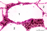
Respiratory bronchiolus (human) | Stain: Hematoxylin and eosin. A longitudinal section shows the lumen of a respiratory bronchiolus (1) with an irregular lining. It is covered by low columnar-cuboidal cells, a few pouches within the same lumen are lined by thin alveolar epithelium as found in the surrounding alveoli (2). Thin arrows... | Respiratory bronchiolus; Columnar epithelium; Cuboidal epithelium; Clara cells; Alveolar epithelium | Poja Histology Collection - Respiratory System Subset |
| 95 |
 |
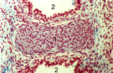
Respiratory bronchiolus (human) | Stain: Azan. The lumen (1) shows an irregular lining. It is covered by low columnar-cuboidal cells (→). Alveolar pouches (*) within the same lumen are lined by thin alveolar epithelium as found in the surrounding alveoli (2). At (5) small patch of smooth muscle, (4) macrophages with phagocytized c... | Respiratory bronchiolus; Columnar epithelium; Cuboidal epithelium; Alveolar epithelium | Poja Histology Collection - Respiratory System Subset |
| 96 |
 |
Respiratory bronchiolus (mouse) | Stain: Hematoxylin and eosin. The lumen (1) shows an irregular lining and is covered by low columnar-cuboidal cells (↓), locally disrupted by a few areas with thin alveolar epithelium as found in the surrounding alveoli (2). Thick arrows point to a row of light-stained Clara cells. At (*) discont... | Respiratory bronchiolus; Columnar epithelium; Cuboidal epithelium; Clara cells; Alveolar epithelium | Poja Histology Collection - Respiratory System Subset |
| 97 |
 |
Respiratory epithelial cells bordering the olfactory region in the nose (human) | Electron microscopy. The five respiratory cells (1) show many cross-sectioned cilia (2) in the lumen. Basal bodies (arrow heads) are regularly arranged at the apex of these cells, and in between, short, slender microvilli. Electron-light cytoplasmic protrusions (3) between the cilia are olfactory bu... | Respiratory epithelium | Poja Histology Collection - Respiratory System Subset |
| 98 |
 |
Respiratory epithelial cells in the nasal concha (rat) | Scanning electron microscopy. Bushes of long cilia (1) are present on the top of the epithelial cells. In between clusters of microvilli-studded goblet cells (2) are visible. | Respiratory epithelium; Pseudostratified epithelium | Poja Histology Collection - Respiratory System Subset |
| 99 |
 |
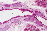
Respiratory epithelium in the nasal septum (rat) | Electron microscopy. At the left pseudostratified epithelium (1) with ciliated columnar (light) cells and electron-dense basal cells (2). In the middle a wide venous capillary (3) with erythrocytes. At the lower part a serous area of a seromucous gland (4) with dark granules and well-developed rough... | Respiratory epithelium; Basal cells | Poja Histology Collection - Respiratory System Subset |
| 100 |
 |
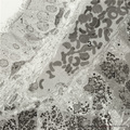
Respiratory epithelium in the nasal vestibulum (rat) | Scanning electron microscopy. Note bushes of cilia (1) on the ciliary epithelium. In between areas of short microvilli-studded bulging or flattened goblet cells. Remnants of erythrocytes (2) are also found on top of ciliated cells. | Respiratory epithelium; Nasal vestibulum | Poja Histology Collection - Respiratory System Subset |