The Health Education Assets Library (HEAL) is a collection of over 22,000 freely available digital materials for health sciences education. The collection is now housed at the University of Utah J. Willard Marriott Digital Library.
TO
Filters: Collection: ehsl_heal
| Title | Description | Subject | Collection | ||
|---|---|---|---|---|---|
| 76 |
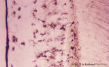 |
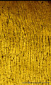
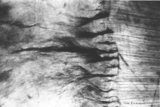
Cellular cementum of the tooth - longitudinal section of root; human, adult. Thin ground section. | Stain: Hematoxylin and eosin. From left to right: remnants of fibers in periodontal ligament (dark band); cementum wih dark lacunae with projecting canaliculi, originally occupied by cementocytes and their processes (broad area); dentinocemental junction; and superficial dentin with S-shaped course ... | oral cavity; cementocytes | Poja Histology Collection - Oral Cavity Subset |
| 77 |
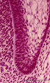 |
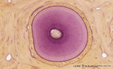
Cervical loop in tooth development - late cap stage, human, embryo | Stain: Azan. From left to right: Dental papilla; Thin basement membrane; Columnar inner dental epithelium (presecretory ameloblasts); Stellate reticulum; Cuboidal outer dental epithelium; Fibrous tooth follicle; Note the transition from inner to outer dental epithelium (cervical loop). | oral cavity; cervical loop | Poja Histology Collection - Oral Cavity Subset |
| 78 |
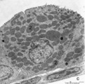 |
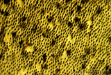
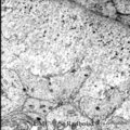
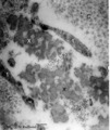
Clara cell in lung (gerbil) | Electron microscopy. In this species a non-ciliated Clara cell (1 = nucleus) contains numerous electron-gray secretion granules (2) of varying sizes, smaller more dense granules tend to localize in the apex of the cell. Between the granules agranular endoplasmic reticulum. Irregular blunt microvill... | Respiratory bronchiolus ; Clara cells; Secretory granules | Poja Histology Collection - Respiratory System Subset |
| 79 |
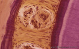 |
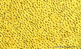
Cross section of tooth in alveolar bone - cat | Stain: Picric acid and hematoxylin. From left to right: alveolar bone covered with a woven type bone (bundle bone) tissue (stained darkly purple); yellow stained periodontal ligament with heavy collagen fibers (horizontal fibers) and blood vessels; bone-related part with Sharpey's fibers followed by... | oral cavity; alveolar bone | Poja Histology Collection - Oral Cavity Subset |
| 80 |
 |
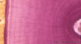
Cross section of tooth in alveolar bone - cat; low magnification | Stain: Picric acid and hematoxylin. From left to right: alveolar bone tissue with osteons; periodontal ligament with blood vessels; acellular cementum (dark purple rim); dentin with (purple) radiair arrangement of dentinal tubules; fine incremental (imbrication) lines of von Ebner run at right angl... | oral cavity; incremental lines; alveolar bone | Poja Histology Collection - Oral Cavity Subset |
| 81 |
 |
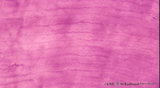
Cross-section of tooth in alveolar bone (cat, adult) | Stain: Picric acid and hematoxylin. From left to right: Periodontal ligament with blood vessels. Acellular cementum (dark purple rim). Dentin with radiair arrangement of dentinal tubules; fine incremental (imbrication) lines of von Ebner run at right angles to these tubules. These lines represent di... | oral cavity; incremental lines; von Ebner | Poja Histology Collection - Oral Cavity Subset |
| 82 |
 |
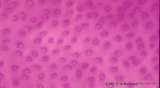
Dentin in cross section of tooth (human, adult) | Stain: Hematoxylin and eosin. Longitudinally sectioned dentinal tubules are parallely arranged, and numerous side branches are visible. | oral cavity; dentinal tubules | Poja Histology Collection - Oral Cavity Subset |
| 83 |
 |
Dentin in cross section of tooth - human, adult | Stain: Hematoxylin and eosin. Cross-sectioned dentinal tubules demonstrate: a light stained center with the odontoblastic process (Tomes' fiber); darker stained peritubular dentin (highly mineralized), also called Neuman's sheath. Intertubular dentin (less mineralized) is present between the tubule... | oral cavity; dentinal tubules; secondary dentin | Poja Histology Collection - Oral Cavity Subset |
| 84 |
 |
Dentinal tubules in longitudinal section of tooth - human, adult. Thin ground section, polarizing microscopy - optical axes of the polarizing plates are crossed at 60. | Slightly oblique section shows dentinal tubules depicted by brown-stained hollow fiber-like structures containing odontoblastic processes (Tomes' fibers) in a semi-three dimensional way. Numerous fine secondary branches of these processes are anastomosing with those of neighboring tubules. | oral cavity; Tomes' Fibers; dentinal tubules | Poja Histology Collection - Oral Cavity Subset |
| 85 |
 |
Dentinal tubules in longitudinal section of tooth - human, adult. Thin ground section, polarizing microscopy - optical axes of the polarizing plates are crossed at 60. | Slightly oblique section shows dentinal tubules depicted by brown-stained hollow fiber-like containing odontoblastic processes (Tomes' fibers) in a semi-three dimensional way ('stubble-field' aspect). Darker stained clusters represent anastomosing secondary branches. | oral cavity; Tomes' Fibers; dentinal tubules | Poja Histology Collection - Oral Cavity Subset |
| 86 |
 |
Dentinal tubules in longitudinal section of tooth - human, adult. Thin ground section, polarizing microscopy - optical axes of the polarizing plates are crossed at 60. | Slightly oblique section shows dentinal tubules depicted by brown-stained stubble structures containing odontoblastic processes (Tomes' fibers) in a semi-three dimensional way. Numerous fine branches of these processes are obvious in dentin. | oral cavity; Tomes' Fibers; dentinal tubules | Poja Histology Collection - Oral Cavity Subset |
| 87 |
 |
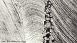
Dentinoenamel junction in longitudinal section of tooth (human, adult). Thin ground section. | From left to right: Enamel with fine striation (course of enamel rods or prisms). Few enamel tufts (left, dark) consisting of hypocalcified enamel rods and interprismatic substance arise from the junction. Scallop-like course of dentinoenamel junction. Dentin with dentinal tubules to the dentinoenam... | oral cavity; enamel tufts; dentinoenamel junction; dentinal tubules | Poja Histology Collection - Oral Cavity Subset |
| 88 |
 |
Dentinoenamel junction in longitudinal section of tooth - human, adult. Thin ground section. | From left to right: enamel with fine striation (composed of stalks of enamel rods or prisms); enamel tufts (dark) arise at the dentinoenamel junction, and these tufts consist of hypocalcified enamel prisms and interprismatic substance; dentin with dentinal tubules up to the dentinoenamel. | oral cavity; enamel tufts; dentinoenamel junction; dentinal tubules | Poja Histology Collection - Oral Cavity Subset |
| 89 |
 |
Dentinoenamel junction in the tooth (human, adult). Thin ground section of crown. | From left to right: Enamel with fine striation (composition of enamel rods or prisms); darker zones almost perpendicular to the striation are the incremental lines (Retzius) due to successive apposition of layers of enamel as the crown is formed. Dentinoenamel junction is shown as a narrow fissure f... | oral cavity; dentinal tubules; dentinoenamel junction; interglobular dentin; lines of Retzius | Poja Histology Collection - Oral Cavity Subset |
| 90 |
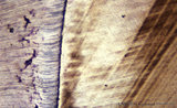 |
Dentinoenamel junction in the tooth - human, adult. Thin ground section of crown. | From left to right: Superficial dentin (bluish) in the crown with S-shaped course of dentinal tubules; They pass uninterrupted through the irregular black structures (due to filling with air) representing hypocalcified areas (interglobular dentin); Mineralization of dentin starts in small globular ... | oral cavity; dentinal tubules; dentinoenamel junction; interglobular dentin; lines of Retzius | Poja Histology Collection - Oral Cavity Subset |
| 91 |
 |
Dentinogenesis in tooth development - bell stage, gerbil, postnatal | Electronmicroscopy. At the top right corner side the distal cytoplasmic parts of presecretory ameloblasts resting on a thin grey basal lamina. In the central area predentin with collagen fibers (grey patches) and cross-sectioned small odontoblastic branches. In between them dispersed numerous dark-s... | oral cavity; predentin; matrix vesicles | Poja Histology Collection - Oral Cavity Subset |
| 92 |
 |
Dentinogenesis in tooth development - bell stage, gerbil, postnatal | Electronmicroscopy. At the bottom side of the predentin partly cross-sectioned odontoblasts with some organelles and many vesicular structures, the dark ones containing hydroxyapatite. Close to the odontoblasts a high concentration of secreted collagen fibers. Further away numerous matrix vesicles (... | oral cavity; predentin; matrix vesicles | Poja Histology Collection - Oral Cavity Subset |
| 93 |
 |
Detail of elastin in an alveolar tip in lung tissue (human, adult) | Electron microscopy. Note the lace-like pattern of lumps of amorph elastin (X) that appears more electron-dense than the collagen fibers (C). At this high magnification cross-sectioned microfibrils (that contain among others fibrillin) are present in close association (*) with different areas of ela... | Elastin-associated proteins; Elastoblast; Myofibroblast | Poja Histology Collection - Respiratory System Subset |
| 94 |
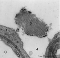 |
Detail of lamellar body (surfactant) and type I alveolar cell in lung (rat) | Electron microscopy. After fixation the extracellular lining of surfactant (phosphatidylcholine, phosphoglycerol, cholesterol and proteins) will often be present as free packed lamellae in the alveolar space (4). This so-called tubular myelin is observed as stacks (5) of lipid crystals and aqueous l... | Pneumocyte I ; Pneumocyte II; Alveolar cells; Tubular myelin | Poja Histology Collection - Respiratory System Subset |
| 95 |
 |
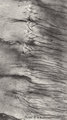
Developing conchae in a frontal section of head (pig, fetus) | Stain: Azan. The fetal conchae develop like a branched gland (1) within the blue-stained trabecular bone (2) of the future skull. The fetal skin (3) is present at the top and bottom. Groups of cheek musculature (4) in middle left and right side. A broad sleeve of chin muscles below (5). The developi... | Conchae nasales; Trabecular bone | Poja Histology Collection - Respiratory System Subset |
| 96 |
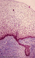 |
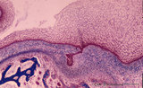
Early cap stage in tooth development - human, embryo | Stain: Azan. From top to bottom: stratified ectoderm with ingrowth of the dental lamina; knob-like end of the dental lamina; and collagen fibers of lamina propria are blue. | oral cavity; tooth development; dental lamina | Poja Histology Collection - Oral Cavity Subset |
| 97 |
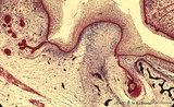 |
Early cap stage in tooth development - human, embryo; low magnification | Stain: Azan. From top to bottom: top left vestibular groove with gland formation; stratified ectoderm with dark red rim of basal cells from which a dental lamina sprouts downwards into the dental crypt (bony cavity) (bottom right corner); bone stains dark blue; connective tissue/mesenchym stains lig... | oral cavity; tooth development; dental lamina | Poja Histology Collection - Oral Cavity Subset |
| 98 |
 |
Early cap stage in tooth development - human, embryo; low magnification | Stain: Azan. From top to bottom: top left vestibular groove with gland formation; stratified ectoderm with ingrowth of the dental lamina (in the middle); bulbous growing end of dental lamina; bottom left alveolar bone formation (dark blue); and connective tissue/mesenchym stains light blue. | oral cavity; tooth development; dental lamina | Poja Histology Collection - Oral Cavity Subset |
| 99 |
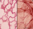 |
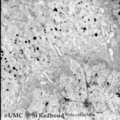
Elastic fibers in the lung (human) | Stain: Orcein. At the left (1) several alveoli (A) are depicted with a faint staining of cellular elements. The elastic fibers within the alveoli are stained reddish-purple. At the right (2) a high magnification of a tangential-sectioned alveolus shows the thicker bundles as well as very thin branch... | Elastic fibers | Poja Histology Collection - Respiratory System Subset |
| 100 |
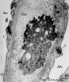 |
Elastin close near the alveolar tip in lung tissue (human, adult) | Electron microscopy. The alveolar tip in the alveolar spaces (Alv) is covered with flattened type I alveolar cells (2). The arrows at (↓1) indicate the junctions between these flattened cells. The amorph elastin (*) in the alveolar tip appears more electron-dense than the collagen fibers (Col). No... | Type I alveolar cells; Elastin-associated proteins | Poja Histology Collection - Respiratory System Subset |