John A. Moran Eye Center Neuro-Ophthalmology Collection: A variety of lectures, videos and images relating to topics in Neuro-Ophthalmology created by faculty at the Moran Eye Center, University of Utah, in Salt Lake City.
NOVEL: https://novel.utah.edu/
TO
| Title | Description | Type | ||
|---|---|---|---|---|
| 76 |
 |
Blepharospasm with Apraxia of the Eye | Image/MovingImage | |
| 77 |
 |
Voluntary Nystagmus | Example of patient with voluntary nystagmus. Discussion of how a lack of uniform, patterned movement of the eyes along with associated lid movements suggests that activity is voluntary. | Image/MovingImage |
| 78 |
 |
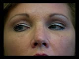
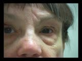
Levator Disinsertion | Example of patient with levator disinsertion, a lid disorder. Patient is pregnant and wears poorly fitting contacts. Discussion of characteristics, such as lid ptosis (shown in the left eye of patient), but with full levator function. | Image/MovingImage |
| 79 |
 |
Marcus Gunn Jaw Winking | Example of patient with Marcus Jaw Winking. Patient is led through instructions for movement of jaw (open, close, back and forth), with eyelid seen to be affected. Patient is then led through instructions for direction of gaze and pursuit. | Image/MovingImage |
| 80 |
 |
CPEO | Patient with Chronic Progressive External Ophthalmoplegia (CPEO) | Image/MovingImage |
| 81 |
 |
Duane's Retraction Syndrome Type 1: Lid Retraction | Example of patients with Duane's Retraction Syndrome, Type 1. Description of components of Duane's Syndrome: limitation of abduction, variable limitation of adduction, and palpebral fissure narrowing and globe retraction with attempted adduction. Type 1 includes limited or absent abduction with norm... | Image/MovingImage |
| 82 |
 |
Cogan's Lid Twitch | Example of a patient with Cogan's lid twitch, with discussion of how to detect it in an exam. | Image/MovingImage |
| 83 |
 |
Fourth Nerve Palsy | Demonstration of examination of patient who experienced blurry vision and pain in the left eye. Demonstrates checking of eye movements, focusing on object while each eye is covered and uncovered, turning head both ways and repeating. Shows limitation of depression in adduction of left eye, left hype... | Image/MovingImage |
| 84 |
 |
3 Step Test | Demonstration of patient examination. | Image/MovingImage |
| 85 |
 |
How to Measure the RAPD | This clip demonstrates the examination technique for measuring the Relative Afferent Pupillary Defect (RAPD). Demonstration of balancing an afferent papillary defect using filters in a patient with a resolving optic neuritis and an afferent papillary defect on the left. | Image/MovingImage |
| 86 |
 |
Duane's Syndrome Type 2: Aberrant Regeneration of the Third and Sixth Nerves | Example of a patient with Type 2 Duane's Syndrome. Demonstrates limitation of adduction in left eye with normal abduction. Discussion of limited pathological cases. | Image/MovingImage |
| 87 |
 |
Duane's Syndrome Type 1 | Clip of patient with Duane's Syndrome Type I. Presented at the Neurology Grand Rounds in Fall 2011 at the University of Utah. Presentation can be found in this collection at: Why Don't You See Double? http://content.lib.utah.edu/u?/EHSL-Moran-Neuro-opth,132 Disease/Diagnosis: Duane's Syndrome Type ... | Image/MovingImage |
| 88 |
 |
Duane's Syndrome Type 3 | Clip of patient with Duane's Syndrome Type III. Presented at the Neurology Grand Rounds in Fall 2011 at the University of Utah. Presentation can be found in this collection at: Why Don't You See Double? http://content.lib.utah.edu/u?/EHSL-Moran-Neuro-opth,132 Disease/Diagnosis: Duane's Syndrome Ty... | Image/MovingImage |
| 89 |
 |
Duane's Syndrome | Example of patient with Duane's Syndrome. Patient is led through instructions for pursuit. | Image/MovingImage |
| 90 |
 |
Measuring Visual Acuity | Demonstration on self of visual acuity exam, using a standard card. | Image/MovingImage |
| 91 |
 |
Testing the Visual Fields | Demonstration of various methods of testing visual fields, including counting fingers, motion, and color of several objects. | Image/MovingImage |
| 92 |
 |
Color Vision Testing | Demonstration of color vision examination. | Text |
| 93 |
 |
Stereoacuity Testing | Demonstration of examination for stereoacuity. | Text |
| 94 |
 |
Amsler Grid Testing | Demonstration of Amsler Grid examination. | Text |
| 95 |
 |
Dilation Lag | Two examples of dilation lag (Horner's syndrome). In the first example, the right pupil dilates much faster than the left pupil when the light is turned out. In the second example, the left pupil dilates much faster than the right pupil when the light is turned out. Discussion of methods of document... | Image/MovingImage |
| 96 |
 |
Parinaud's Syndrome | Two examples of patients with Parinaud's syndrome, a dorsal midbrain syndrome. Discussion of hallmarks of this syndrome, including convergence retraction nystagmus, vertical gaze palsies, light-near dissociation, and Collier's Sign. Discussion of age-dependent disorders associated with this syndrome... | Image/MovingImage |
| 97 |
 |
Sector Palsies and Light-Near Dissociation | Example of patient with bilateral Adie's pupils. Exam is performed with a slit-lamp. Shows iris stroma and focal segments of iris sphincter that retain their contractilty. Suggests post-ganglionic parasympathetic denervation. | Image/MovingImage |
| 98 |
 |
RAPD Present | This clip demonstrates the technique used to determine that Relative Afferent Pupillary Defect (RAPD) is present in a patient. | Image/MovingImage |
| 99 |
 |
Normal Light Reflex without RAPD | This clip demonstrates the examination of the Relative Afferent Pupillary Defect (RAPD.) Demonstration of gauging the size of the pupil in light, testing light reflexes, swinging flashlight test for optic nerve abnormality. | Image/MovingImage |
| 100 |
 |
Pupil Exam | Demonstration of pupil examination. | Text |