The Health Education Assets Library (HEAL) is a collection of over 22,000 freely available digital materials for health sciences education. The collection is now housed at the University of Utah J. Willard Marriott Digital Library.
TO
| Title | Description | Subject | Collection | ||
|---|---|---|---|---|---|
| 76 |
 |
Diagram: type I vs. type II 2nd degree AV block | In type I 2nd degree AV block the PR progressively lengthens until a nonconducted P wave occurs. The PR gets longer by smaller and smaller increments; this results in gradual shortening of the RR intervals. The RR interval of the pause is usually less than the two preceding RR intervals. The RR i... | Knowledge Weavers ECG | |
| 77 |
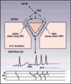 |
Diagram: AV nodal reentrant tachycardia | The AV node often has dual pathways; in this diagram the alpha pathway is fast, but has a long refractory period; the beta pathway is conducts more slowly, but recovers faster.In sinus rhythm the faster alpha pathway is used and accounts for the normal PR interval. When a PAC occurs, however, the i... | Knowledge Weavers ECG | |
| 78 |
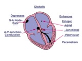 |
Diagram: digitalis effect on rhythm and conduction | Diagram: digitalis effect on rhythm and conduction | Knowledge Weavers ECG | |
| 79 |
 |
Diffuse anterolateral T wave abnormalities | Diffuse anterolateral T wave abnormalities | T Wave Abnormalities | Knowledge Weavers ECG |
| 80 |
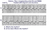 |
Digitalis intoxication: Junctional tachycardia with and without AV block | In a patient with longstanding atrial fibrillation being treated with digoxin, a regular tachycardia, as seen in A, with a RBBB suggests a junctional or supraventricular tachycardia. Group beating, in B, is likely due to a 2nd degree, Type 1, exit block below the ectopic junctional focus. This is h... | Knowledge Weavers ECG | |
| 81 |
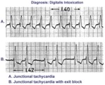 |
Digitalis intoxication: junctional tachycardia with and without exit block | In A the rhythm is junctional tachycardia with RBBB. In B there is 2nd degree exit block with a 3:2 conduction ratio; i.e., every 3rd junctional impulse fails to reach the ventricles... at least for the first two groupings on 1.4sec. | Knowledge Weavers ECG | |
| 82 |
 |
ECG components diagram - marquette | ECG components diagram - marquette | Knowledge Weavers ECG | |
| 83 |
 |
ECG intervals and waves | The P wave represents atrial activation; the PR interval is the time from onset of atrial activation to onset of ventricular activation. The QRS complex represents ventricular activation; the QRS duration is the duration of ventricular activation. The ST-T wave represents ventricular repolarizatio... | Knowledge Weavers ECG | |
| 84 |
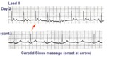 |
ECG of the century - part II: dual AV pathways | An astute cardiology fellow, yours truly, went to the patient's bedside on Day 2 and massaged the right carotid sinus as indicated by the arrow. Four beats later at a slightly slower heart rate the PR interval suddenly normalized suggesting an abrupt change from a slow AV nodal pathway to a fast AV... | Knowledge Weavers ECG | |
| 85 |
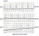 |
ECG of the century: A most unusual 1st degree AV block | On Day 1, at a heart rate of 103 bpm the P waves are not clearly defined suggesting an accelerated junctional rhythm. However, on Day 2, at a slightly slower heart rate the sinus P wave suddenly appears immediately after the QRS complex. In retrospect, the sinus P wave in Day 1 was found burried i... | Knowledge Weavers ECG | |
| 86 |
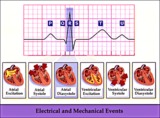 |
Electrical and mechanical events diagram - marquette | Electrical and mechanical events diagram - marquette | Knowledge Weavers ECG | |
| 87 |
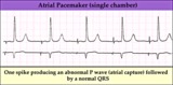 |
Electronic atrial pacing - marquette | Electronic atrial pacing - marquette | Knowledge Weavers ECG | |
| 88 |
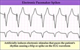 |
Electronic ventricular pacemaker rhythm - marquette | Electronic ventricular pacemaker rhythm - marquette | Knowledge Weavers ECG | |
| 89 |
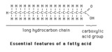 |
Essential features of a fatty acid | The essential features of a fatty acid are a long hydrocarbon chain terminating in a carboxylic acid group. | Knowledge Weavers Fatty Acids | |
| 90 |
 |
Extensive anterior/anterolateral MI: precordial leads | Extensive anterior/anterolateral MI: precordial leads | Knowledge Weavers ECG | |
| 91 |
 |
Extensive anterior/anterolateral MI: recent | Significant pathologic Q-waves (V2-6, I, aVL) plus marked ST segment elevation are evidence for this large anterior/anterolateral MI. The exact age of the infarction cannot be determined without clinical correlation and previous ECGs, but this is likely a recent MI. | Knowledge Weavers ECG | |
| 92 |
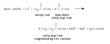 |
Fatty acid elongation in mitochondria | This shows the overall reaction of fatty acid elongation in mitochondria. The process is essentially a reversal of beta-oxidation, except that one NADPH and one NADH are required (beta-oxidation yields two NADH). Mitochondrial fatty acid elongation acts primarily on fatty acyl CoA substrates short... | Knowledge Weavers Fatty Acids | |
| 93 |
 |
Fatty acid metabolism -- schematic overview | Fatty acids are taken up by cells, where thy may serve as precursors in the synthesis of other compounds, as fuels for energy production, and as substrates for ketone body synthesis. Ketones bodies may then be exported to other tissues, where they can be used for energy production. | Biosynthesis | Knowledge Weavers Fatty Acids |
| 94 |
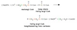 |
Fatty acyl CoA elongation in the endoplasmic reticulum | This shows the overall process of fatty acyl elongation in the endoplasmic reticulum. The process resembles that catalyzed by fatty acyl synthase, but the individual activities appear to be on separate enzymes. | Knowledge Weavers Fatty Acids | |
| 95 |
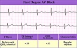 |
First degree AV block - marquette | First degree AV block - marquette | Knowledge Weavers ECG | |
| 96 |
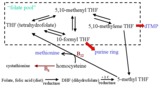 |
Folate Pool - The Role of B Vitamins in One-Carbon Metabolism | This figure depicts the pathway for folate utilization and the role of vitamins B6 and B12 in the metabolism of methyl-tetrahydrofolate and homocysteine. | Folate; Folicin | HEAL Open Review Collection |
| 97 |
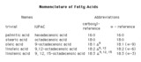 |
Four systems for denoting fatty acids | There are four commonly used ways of designating fatty acids. The first two columns show samples of names, and the last two columns show systems of abbreviating these names. | Nomenclature | Knowledge Weavers Fatty Acids |
| 98 |
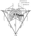 |
Frontal and horizontal plane lead diagram | Frontal and horizontal plane lead diagram | Knowledge Weavers ECG | |
| 99 |
 |
Frontal plane QRS axis = +15 degrees | Frontal plane QRS axis = +15 degrees | Knowledge Weavers ECG | |
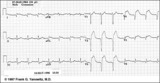
| 100 |
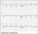 |
Frontal plane QRS axis = +150 degrees (RAD) | This is an unusual right axis deviation (RAD). Lead I is negative, which usually means RAD. Lead II is the isoelectric lead, which almost always means -30 degrees; but in this example the axis is 180 degrees away from -30, or +150 degrees. | Knowledge Weavers ECG |