John A. Moran Eye Center Neuro-Ophthalmology Collection: A variety of lectures, videos and images relating to topics in Neuro-Ophthalmology created by faculty at the Moran Eye Center, University of Utah, in Salt Lake City.
NOVEL: https://novel.utah.edu/
TO
| Title | Description | Type | ||
|---|---|---|---|---|
| 76 |
 |
Duane's Syndrome | Example of patient with Duane's Syndrome. Patient is led through instructions for pursuit. | Image/MovingImage |
| 77 |
 |
Measuring Visual Acuity | Demonstration on self of visual acuity exam, using a standard card. | Image/MovingImage |
| 78 |
 |
Testing the Visual Fields | Demonstration of various methods of testing visual fields, including counting fingers, motion, and color of several objects. | Image/MovingImage |
| 79 |
 |
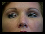
Dilation Lag | Two examples of dilation lag (Horner's syndrome). In the first example, the right pupil dilates much faster than the left pupil when the light is turned out. In the second example, the left pupil dilates much faster than the right pupil when the light is turned out. Discussion of methods of document... | Image/MovingImage |
| 80 |
 |
Parinaud's Syndrome | Two examples of patients with Parinaud's syndrome, a dorsal midbrain syndrome. Discussion of hallmarks of this syndrome, including convergence retraction nystagmus, vertical gaze palsies, light-near dissociation, and Collier's Sign. Discussion of age-dependent disorders associated with this syndrome... | Image/MovingImage |
| 81 |
 |
Sector Palsies and Light-Near Dissociation | Example of patient with bilateral Adie's pupils. Exam is performed with a slit-lamp. Shows iris stroma and focal segments of iris sphincter that retain their contractilty. Suggests post-ganglionic parasympathetic denervation. | Image/MovingImage |
| 82 |
 |
RAPD Present | This clip demonstrates the technique used to determine that Relative Afferent Pupillary Defect (RAPD) is present in a patient. | Image/MovingImage |
| 83 |
 |
Transillumination - Lisch Nodules | Demonstration of transillumination of the Lisch nodules on a patient with neurofibromatosis. Shows how Lisch nodules that were not very visible in slit-lamp examination are better seen with transillumination, which may therefore be useful in detecting Lisch nodules earlier in children where they are... | Image/MovingImage |
| 84 |
 |
Transillumination - Ciliary Body Neurofibromas | Example of transillumination on a patient with neurofibromatosis, but without Lisch nodules. Shows suspected neurofibromas in the ciliary body. | Image/MovingImage |
| 85 |
 |
How to Use the Direct Ophthalmoscope in an Exam | Demonstration of using the direct ophthalmoscope to examine the optic disc. Covers hand placement , which eye to use, and distance from patient. | Image/MovingImage |
| 86 |
 |
Tour of the Fundus | This clip demonstrates the funduscopic examination technique. | Image/MovingImage |
| 87 |
|
Progressive Supranuclear Palsy | Example of patient with progressive supranuclear palsy. Discussion of difference between saccadic movement in supranuclear palsy and nystagmus. Shows saccadic intrusions in forward gaze, pursuit, saccades, and doll's head maneuver. | Image/MovingImage |
| 88 |
 |
Before Tensilon | Example of patient with myasthenia gravis. Demonstration of baseline examination, followed by administration of 2mg of tensilon, which is a test dose. Procedure for administration of tensilon test is described, including variations. Patient is then shown after being given 4mg of tensilon, with very ... | Image/MovingImage |
| 89 |
 |
Tour of the Direct Ophthalmoscope | This clip describes the parts and operation of the ophthalmoscope as an ocular examination tool. Includes adjustment of aperture size and adjustment of lenses. | Image/MovingImage |
| 90 |
 |
Basic Eye Alignment Exam | Demonstration of basic eye alignment examination. Includes: a. Tools b. Cover-Uncover and SPCT c. Alternate Cover and APCT d. Maddox Rod Testing | Image/MovingImage |
| 91 |
 |
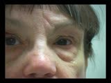
Bilateral Asynchronous Blepharospasm with Facial and Cervical Dystonia | Bilateral Asynchronous Blepharospasm with Facial and Cervical Dystonia. | Image/MovingImage |
| 92 |
 |
Normal Light Reflex without RAPD | This clip demonstrates the examination of the Relative Afferent Pupillary Defect (RAPD.) Demonstration of gauging the size of the pupil in light, testing light reflexes, swinging flashlight test for optic nerve abnormality. | Image/MovingImage |
| 93 |
 |
See-saw Nystagmus | 7-year-old female whose mother noticed her eyes "bouncing" for 2 months. Visual acuity 20/70 OD and 20/40 OS, reduced color vision OU, and no afferent pupillary defect. See-saw nystagmus documented with videography. Manual perimetry revealed a complete right homonymous hemianopia. MRI revealed a lar... | Image/MovingImage |
| 94 |
 |
Cyclic Oculomotor Palsy | Example of patient with cyclic oculomotor palsy. | Image/MovingImage |
| 95 |
 |
Progressive Supranuclear Palsy | Progressive Supranuclear Palsy | Image/MovingImage |
| 96 |
 |
Oculopalatal Myoclonus (PPT) | Oculopalatal myoclonus (OPM) Rhythmic oscillations of eyes and palate. Occurred after specific brainstem injury from stroke, following stenting. Related Video: http://content.lib.utah.edu/u?/EHSL-Moran-Neuro-opth,128 Disease/Diagnosis: Oculopalatal myoclonus | Image/MovingImage |
| 97 |
 |
Oculopalatal Myoclonus | Oculopalatal myoclonus (OPM) Rhythmic oscillations of eyes and palate. Occurred after specific brainstem injury from stroke, following stenting. Related PowerPoint Presentation: http://content.lib.utah.edu/u?/EHSL-Moran-Neuro-opth,129 Disease/Diagnosis: Oculopalatal myoclonus. | Image/MovingImage |
| 98 |
 |
Duane's Syndrome Type 1 | Clip of patient with Duane's Syndrome Type I. Presented at the Neurology Grand Rounds in Fall 2011 at the University of Utah. Presentation can be found in this collection at: Why Don't You See Double? http://content.lib.utah.edu/u?/EHSL-Moran-Neuro-opth,132 Disease/Diagnosis: Duane's Syndrome Type ... | Image/MovingImage |
| 99 |
 |
Duane's Syndrome Type 3 | Clip of patient with Duane's Syndrome Type III. Presented at the Neurology Grand Rounds in Fall 2011 at the University of Utah. Presentation can be found in this collection at: Why Don't You See Double? http://content.lib.utah.edu/u?/EHSL-Moran-Neuro-opth,132 Disease/Diagnosis: Duane's Syndrome Ty... | Image/MovingImage |
| 100 |
 |
A-scan Technique | This video describes and demonstrates the A-scan examination technique for examination of the eye using ultrasonography. | Image/MovingImage |