The Health Education Assets Library (HEAL) is a collection of over 22,000 freely available digital materials for health sciences education. The collection is now housed at the University of Utah J. Willard Marriott Digital Library.
TO
| Title | Description | Subject | Collection | ||
|---|---|---|---|---|---|
| 76 |
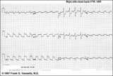 |
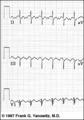
Acute infero-postero-lateral MI | Hyperacute ST segment elevation is seen in leads II, III, aVF (inferior location) and in leads V4-6 (apical lateral wall location). Hyperacute ST depression is seen in leads V1-2 (an expression of posterior wall injury). in addition there are reciprocal ST segment depression changes in leads I an... | Knowledge Weavers ECG | |
| 77 |
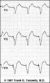 |
Ventricular Pacemaker Rhythm: V1-3 | Note the small pacemaker spikes before the QRS complexes. In addition, the QRS complex in V1-3 exhibits ventricular ectopic morphology; i.e., there is a slur or notch at the beginning of the S wave, and >60ms delay from onset to QRS to nadir of S wave. This rules against a supraventricular rhythm wi... | Knowledge Weavers ECG | |
| 78 |
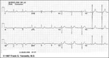 |
Inferior MI: fully evolved | Significant pathologic Q-waves are seen in leads II, III, aVF along with resolving ST segment elevation and symmetrical T wave inversion. This is a classic inferior MI. | Knowledge Weavers ECG | |
| 79 |
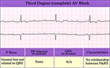 |
Complete AV block (3rd degree) with junctional rhythm | Complete AV block (3rd degree) with junctional rhythm | Knowledge Weavers ECG | |
| 80 |
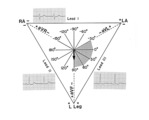 |
QRS axis = +90 degrees | Lead I is isoelectric; II and III are positive; the axis is +90 degrees. | Knowledge Weavers ECG | |
| 81 |
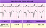 |
Left bundle branch block - marquette | Left bundle branch block - marquette | Knowledge Weavers ECG | |
| 82 |
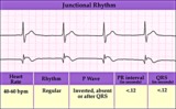 |
Junctional escape rhythm | Junctional escape rhythm | Knowledge Weavers ECG | |
| 83 |
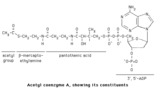 |
Acetyl CoA structure | Acetyl CoA Structure. The thioester bond linking the acetyl group to CoA is ahigh energybond. | Knowledge Weavers Fatty Acids | |
| 84 |
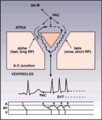 |
Diagram: AV nodal reentrant tachycardia | The AV node often has dual pathways; in this diagram the alpha pathway is fast, but has a long refractory period; the beta pathway is conducts more slowly, but recovers faster.In sinus rhythm the faster alpha pathway is used and accounts for the normal PR interval. When a PAC occurs, however, the i... | Knowledge Weavers ECG | |
| 85 |
 |
Atrial parasystole | In atrial parasystole non-fixed coupled PACs, shown by arrows, occur at a common inter-ectopic interval or at multiples of this interval. Atrial fusions, not shown here, may also occur when the PAC occurs in close temporal proximity to the sinus impulse. | Knowledge Weavers ECG | |
| 86 |
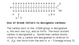 |
Use of greek letters to designate carbons in fatty acids | The carbon next to the -COOH group is the alpha carbon; the next one is the beta carbon, and so forth. The carbon most distant from the -COOH group is designated omega. Carbons close to the omega carbon may be designated in relation to it. E.g., the third carbon from the end is omega - 3, and the... | Nomenclature | Knowledge Weavers Fatty Acids |
| 87 |
 |
Atrial flutter with 2:1 conduction: leads II, III, V1 | Atrial flutter with 2:1 conduction: leads II, III, V1 | Knowledge Weavers ECG | |
| 88 |
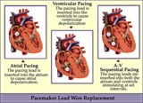 |
Pacemaker lead wire placement diagram - marquette | Pacemaker lead wire placement diagram - marquette | Knowledge Weavers ECG | |
| 89 |
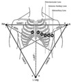 |
Frontal and horizontal plane lead diagram | Frontal and horizontal plane lead diagram | Knowledge Weavers ECG | |
| 90 |
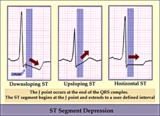 |
ST segment diagram - marquette | ST segment diagram - marquette | Knowledge Weavers ECG | |
| 91 |
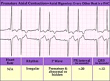 |
Atrial bigeminy - marquette | Atrial bigeminy - marquette | Knowledge Weavers ECG | |
| 92 |
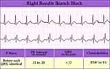 |
Right Bundle Branch Block | Right Bundle Branch Block | Knowledge Weavers ECG | |
| 93 |
 |
2nd degree AV block, type I, with accelerated junctional escapes and a ladder diagram | The ladder diagram illustrates a Wenckebach type AV block by the increasing PR intervals before the blocked P wave. After the blocked P wave, however, a rev-ed up junctional pacemaker terminates the pause. Note that the junctional beats have a slightly different QRS morphology from the sinus beats... | Knowledge Weavers ECG | |
| 94 |
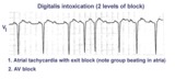 |
Atrial tachycardia with exit block and AV block | The ectopic P waves, easily seen in this example,occur in groups, separated by short pauses. This is likely due to an exit block just distal to the atrial pacemaker. Because not all of the P waves make it to the ventricles, there is also 2nd degree AV block. Therefore, two levels of block are pre... | Knowledge Weavers ECG | |
| 95 |
 |
LVH | In this example of LVH, the precordial leads don't meet the usual voltage criteria or exhibit significant ST segment abnormalities. The frontal plane leads, however, show voltage criteria for LVH and significant ST segment depression in leads with tall R waves. The voltage criteria include 1) R in a... | Knowledge Weavers ECG | |
| 96 |
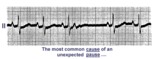 |
A nonconducted PAC causes an unexpected pause | Unexpected pauses in rhythm have several causes, the most frequent being a nonconducted PAC. In this example the nonconducted PAC is seen in the ST segment of the pause. Note the change in the ST-T compared to the other ST-T waves. | Knowledge Weavers ECG | |
| 97 |
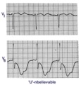 |
Giant TU fusion waves | TU fusion waves are often seen in long QT syndromes. The differential diagnosis of this ECG abnormality includes electrolyte abnormalities -hypokalemia, CNS disease, e.g., subarachnoid hemorrhage; hereditary long QT syndromes, and drugs such as quinidine. | Knowledge Weavers ECG | |
| 98 |
 |
Left anterior fascicular block (LAFB) | LAFB is the most common of the intraventricular conduction defects. It is recognized by 1) left axis deviation; 2) rS complexes in II, III, aVF; and 3) small q in I and/or aVL. | Knowledge Weavers ECG | |
| 99 |
 |
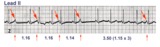
Atrial flutter with 2:1 AV conduction: lead V1 | The arrows point to two flutter waves for each QRS complex. Atrial rate = 280; ventricular rate = 140. | Knowledge Weavers ECG | |
| 100 |
 |
Long QT Interval and Giant Negative T Waves | Long QT Interval and Giant Negative T Waves | Knowledge Weavers ECG |