The Health Education Assets Library (HEAL) is a collection of over 22,000 freely available digital materials for health sciences education. The collection is now housed at the University of Utah J. Willard Marriott Digital Library.
TO
Filters: Collection: ehsl_heal
| Title | Description | Subject | Collection | ||
|---|---|---|---|---|---|
| 51 |
 |
Basophilic granulocyte (peripheral blood, human) | Electron microscopy. The detail shows close to the nucleus characteristic developing basophilic granules (specific granules) (8-13 μm). At thin arrow (↓) a small Golgi area with a small specific granule. At (***) granules with different osmiophilic internal structures. The basophilic granules var... | Poja Histology Collection - Blood & Bone Marrow Subset | |
| 52 |
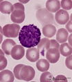 |
Basophilic granulocyte in peripheral blood smear (human) | Stain: May-Grnwald-Giemsa (MGG). The basophilic granulocyte has large, coarse dark purple-stained granules that contain mediators such as eosinophilic chemotactic factors, heparin, histamine, myeloperoxidase. The nuclear lobes are irregular, badly visible and covered by the granules. | Poja Histology Collection - Blood & Bone Marrow Subset | |
| 53 |
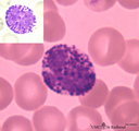 |
Basophilic granulocyte in peripheral blood smear (human) | Stain: May-Grnwald-Giemsa (MGG). The basophilic granulocyte is characterized by large, coarse, aggregated dark purple granules. The nuclear lobes are usually not very well visible and masked by the granules (see also inset). | Poja Histology Collection - Blood & Bone Marrow Subset | |
| 54 |
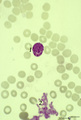 |
Basophilic myelocyte in bone marrow smear (human) | Stain: May-Grnwald-Giemsa (MGG). The basophilic myelocyte (1) contains dark bluish-purple (metachromatic), coarse granules in the cytoplasm. (2) Platelets. | Poja Histology Collection - Blood & Bone Marrow Subset | |
| 55 |
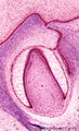 |
Bell stage of the tooth development - human, embryo; low magnification | Stain: Azan. From top to bottom: Stratified ectoderm with a distinct basal layer (red line) of cuboid cells; Dental lamina giving rise to the bell stage (left) and to the primordium of permanent tooth (right); Odontogenic organ (future deciduous tooth) surrounded by fibrous tooth follicle; Out... | oral cavity; dental lamina; predentin | Poja Histology Collection - Oral Cavity Subset |
| 56 |
 |
Binucleated plasma cell in peripheral blood smear (human) | Stain: May-Grnwald-Giemsa (MGG). A large plasma cell with two nuclei. The chromatin is coarsely clumped. The cytoplasm is intensive basophilic due to the large RER content for purpose of antibody production. Note the clear zone (Golgi area) between the nuclei. | Poja Histology Collection - Blood & Bone Marrow Subset | |
| 57 |
 |
Border of a marginal zone in spleen (rat) | Stain: Immunoelectron microscopy (gold labeling of heparan sulfate in Lowicryl embedding, using the single chain antibody HS4C3). The zone of red pulp immediately surrounding a lymphatic nodule is called the marginal zone (perilymphoid zone) and is composed of a scaffold of basal lamina material w... | marginal zone; immuno electron microscopy; heparan sulfate; dendritic cell | Poja Histology Collection - Lymphatic Tissues and Organs Subset |
| 58 |
 |
Border of white pulp in spleen (mouse) | Monoaminooxidase (enzyme histochemistry on frozen section) with Nitro-BT as staining substrate resulting in a blue formazan precipitate. Despite the general activity of most cells in the spleen, the border cells or so-called metallophilic cells (1) or dendritic antigen-presenting cells (APC) show th... | monoamino oxidase; ED3 antibody; follicle; white pulp | Poja Histology Collection - Lymphatic Tissues and Organs Subset |
| 59 |
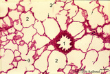 |
Bronchiolus and alveoli in lung (human, adult) | Stain: Hematoxylin and eosin. (1) indicates a bronchiolus with respiratory epithelium and muscle fibers in its wall. Alveolar spaces (2) are separated by alveolar cell types that lined thin septa with capillaries. The septa end in tips and knobs (3). | Bronchiolus; Alveolar septa; Pneumocyte I; Pneumocyte II; Alveolar cell types | Poja Histology Collection - Respiratory System Subset |
| 60 |
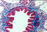 |
Bronchiolus in the lung (human, adult) | Stain: Azan. The lumen of the bronchiolus is lined with one layer of ciliated epithelium (1) and is folded due to the contraction of smooth muscle fibers (2) in the wall. Note the rich cellularity (3) in the stromal surrounding of the bronchiolus. Alveolar space (4). (5) pulmonary artery branches. | Ciliated epithelium; Bronchiolus | Poja Histology Collection - Respiratory System Subset |
| 61 |
 |
Bronchiolus in the lung (human, adult) | Stain: Azan. The lumen is lined with one layer of ciliated epithelium (1) without goblet cells. Patches of smooth muscle fibers (2) are present. Notice small bronchial arteries (*). Arrows (↓) indicate sites of carbon accumulations between the alveoli (3). | Bronchiolus; Ciliated epithelium | Poja Histology Collection - Respiratory System Subset |
| 62 |
 |
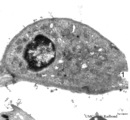
Brush cell in lung (rat) | Electron microscopy. In the proximal as well as in the terminal airways a special type of cell the so-called brush(-border) cell could be observed. In this picture this cell (X) is located in the alveolar space. At (*) the brush border, the dense cytoplasm contains many organelles. (C) is a capillar... | Brush cells | Poja Histology Collection - Respiratory System Subset |
| 63 |
 |
Brush cell in lung (rat) | Electron microscopy. In the proximal as well as in the terminal airways a special type of cell can be observed, the so-called brush (-border) cell. In this picture this cell is located in the alveolar space. At (1) the brush border of thick plump microvilli. In the cytoplasm a Gogi area, mitochondri... | Brush cell | Poja Histology Collection - Respiratory System Subset |
| 64 |
 |
Bud stage outgrowth in tooth development - tooth germ, human, embryo | Stain: hematoxylin. At the top stratified ectoderm continuous with the former epithelial tooth bud that abuts toward the underlying neural crest-derived mesenchymal cells. A basal lamina delimits the palisade-arranged epithelial cells from the surrounding dense aggregate of mesenchymal cells. | oral cavity; tooth bud; tooth development | Poja Histology Collection - Oral Cavity Subset |
| 65 |
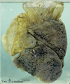 |
Bullous emphysema of the lung (honeycomb lung, human, adult) | Macroscopy of the left lung showing large apical bullae (3) due to total destruction of all alveoli leaving remnants of the covering pleura. (2) indicates small bullae of foci of destroyed lobules with permanent enlargement of the air spaces and (1) points to spots of anthracosis in less-affected lu... | Macroscopy; Honeycomb lung; Interstitial fibrosis; Hamman-Rich Syndrome | Poja Histology Collection - Respiratory System Subset |
| 66 |
 |
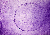
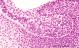
Calcified bacterial plaque in palatine tonsil ('gut-associated lymphatic tissue' or GALT) (human) | Stain: Azan. A tonsillar crypt lined by squamous epithelium (2) that is infiltrated with lymphocytes (1). It is a normal finding that within the crypts free cells, plugs of lymphocytes (1) and calcified epithelial debris as well as colonies of oral commensally bacteria are present. (3) shows a calc... | bacterial plaque | Poja Histology Collection - Lymphatic Tissues and Organs Subset |
| 67 |
 |
Capillary system of a lung alveolus (cat) | Stain: specimen injected with India ink via the pulmonary system. In an en face view of the alveolar wall the black- and granular-stained capillary plexus is well shown. The larger vessels represent branches of the pulmonary arteriole. | Poja Histology Collection - Respiratory System Subset | |
| 68 |
 |
Capillary system of an alveolus (cat) | Stain: specimen injected with trypan blue in gelatin via the pulmonary system. In a tangential view the blue-stained dense capillary plexus of one alveolus is well shown. | Poja Histology Collection - Respiratory System Subset | |
| 69 |
 |
Capillary system of lung alveoli (cat) | Stain: specimen injected with trypan blue in gelatin via the pulmonary system. In a tangential view the blue-stained dense capillary plexus surrounding each alveolus is well depicted. | Poja Histology Collection - Respiratory System Subset | |
| 70 |
 |
Cellular cementum of the tooth - longitudinal section of root; human, adult. Thin ground section. | Stain: Hematoxylin and eosin. From left to right: remnants of fibers in periodontal ligament (dark band); cementum wih dark lacunae with projecting canaliculi, originally occupied by cementocytes and their processes (broad area); dentinocemental junction; and superficial dentin with S-shaped course ... | oral cavity; cementocytes | Poja Histology Collection - Oral Cavity Subset |
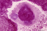
| 71 |
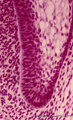 |
Cervical loop in tooth development - late cap stage, human, embryo | Stain: Azan. From left to right: Dental papilla; Thin basement membrane; Columnar inner dental epithelium (presecretory ameloblasts); Stellate reticulum; Cuboidal outer dental epithelium; Fibrous tooth follicle; Note the transition from inner to outer dental epithelium (cervical loop). | oral cavity; cervical loop | Poja Histology Collection - Oral Cavity Subset |
| 72 |
 |
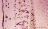
Chorioamnionitis (human) | Histologically chorioamnionitis describes the progression of the inflammatory process. Bacteria firstly colonized the chorioamniotic surface. In first two days polymorphonuclear granulocytes (PMN) migrate to the chorion (chorionitis) marginate and adhere to the bottom of the chorionic plate (stage 1... | chorioamnionitis; placenta; trophoblast | Poja Histology Collection - Placenta |
| 73 |
 |
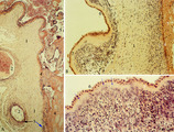
Chorioangioma (human) | Stain: (A) Hematoxylin-eosin (survey) and (B, C) van Gieson. (A) chorioangioma (1), chorionic plate (2), stem villus (3) and villi (5). (B) shows in (1) a large cellular mass with accumulated capillaries. At (2) part of the chorionic plate. Subchorionic fibrinoid stains yellow (4). (C) reveals t... | placenta; chorioangioma; villus; chorionic plate | Poja Histology Collection - Placenta |
| 74 |
 |
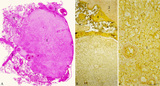
Choriocarcinoma (human) | Stain: Hematoxylin-eosin. (A) Inset: Survey tumor. Within the uterus (1) a choriocarcinoma forms solitary or multiple nodules composed of hemorrhagic necrotic areas (2) surrounded by neoplastic cells. It resembles an early implanted blastocyst with aggregations of mononuclear lighter-stained cyt... | choriocarcinoma; placenta; uterus; cytotrophoblast; GTD (gestational trophoblastic disease) | Poja Histology Collection - Placenta |
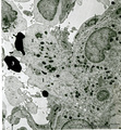
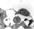
| 75 |
 |
Circulating phagocytic cells | Electron microscopy, a set of the ultrastuctural features of different types of phagocytic cells. Top left side (A): Circulating monocyte with phagocytized latex particles (peripheral blood, human). The nucleus is large and with a long small indentation. Close to the nucleus several individually in... | Poja Histology Collection - Blood & Bone Marrow Subset |