The Health Education Assets Library (HEAL) is a collection of over 22,000 freely available digital materials for health sciences education. The collection is now housed at the University of Utah J. Willard Marriott Digital Library.
TO
Filters: Collection: ehsl_heal
| Title | Description | Subject | Collection | ||
|---|---|---|---|---|---|
| 51 |
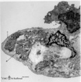 |
The alveolar tip in lung tissue (human, adult) | Electron microscopy. The lung tip is covered by a flattened alveolar cell type I (↓1) and a similar neighboring bulging one with nucleus (2). In the alveolar tip amorph elastin (5) appears more electron-dense than the collagen fibers (Col) and is interwoven with it. Small foci of calcifications a... | Alveolar tip; Elastoblast | Poja Histology Collection - Respiratory System Subset |
| 52 |
 |
The alveolus and air-blood barrier in the lung (rat) | Electron microscopy. The alveolus (1) is lined by a thin extension (2) of the alveolar epithelial cell type I (2), the pneumocyte I and the thin endothelium (3) of the capillary filled with erythrocytes (4) and blood platelets (5). The thin-walled air-blood barrier (↔) consists of the transition f... | Pneumocyte I; Alveolar cell type I | Poja Histology Collection - Respiratory System Subset |
| 53 |
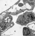 |
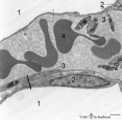
Alveolus in lung (dog) | Electron microscopy. (A) indicate alveolar space; (C) indicate capillary with erythrocyte. Drawing-pin represents endothelium. (1) type I alveolar cell; (↓) indicate thin cytoplasm of pneumocyte I. (2) type II alveolar cell. (3) alveolar macrophage. (*) are spots of elastin, intermingled with coll... | Pneumocyte I; Pneumocyte II; Alveolar cells; Alveolar macrophage | Poja Histology Collection - Respiratory System Subset |
| 54 |
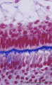 |
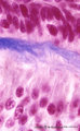
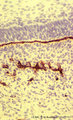
Ameloblasts and odontoblasts in tooth development - advanced bell stage, human, embryo | Stain: Azan. From top to bottom: Stellate reticulum consisting of a loose network of ectoderm-derived cells; Cell layers of the stratum intermedium; Columnar presecretory ameloblasts with their nuclear area close to the stratum intermedium, and at the distal side (secretion area) oriented towards pr... | oral cavity; predentin | Poja Histology Collection - Oral Cavity Subset |
| 55 |
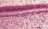 |
Ameloblasts and odontoblasts in tooth development - advanced bell stage, human, embryo | Stain: Hematoxylin and eosin. From top to bottom: Stellate reticulum consisting of a loose network of ectoderm-derived cells; Cell layers of the stratum intermedium; Palisade-arranged columnar presecretory ameloblasts at the distal side oriented towards the basal lamina enforced by deposited collage... | oral cavity | Poja Histology Collection - Oral Cavity Subset |
| 56 |
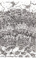 |
Ameloblasts and odontoblasts in tooth development - advanced bell stage, human, embryo CR 80 mm | Scheme electronmicroscopy. From top to bottom: Stellate reticulum consisting of a non-vascularized network of ectoderm-derived cells continuous with the cell layers of the stratum intermedium; Columnar presecretory ameloblasts with their upper side (nuclear area) in close contact with the stratum in... | oral cavity; predentin | Poja Histology Collection - Oral Cavity Subset |
| 57 |
 |
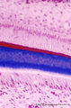
Ameloblasts and odontoblasts in tooth development - advanced bell stage, human, embryo; high magnification | Stain: Azan. From top to bottom: A cell layer of the stratum intermedium; Columnar presecretory ameloblasts at the distal side (secretion area) oriented towards predentin (blue); Note a gradient from left to right in the amount of deposited collagen fibers; Tall columnar odontoblasts in an epithelio... | oral cavity | Poja Histology Collection - Oral Cavity Subset |
| 58 |
 |
Ameloblasts and odontoblasts in tooth development - bell stage, human, embryo | Stain: Azan. From top to bottom: Stellate reticulum consisting of a loose network of ectoderm-derived cells; Cell layers of the stratum intermedium; Palisade-arranged tall columnar secretory ameloblasts with their nuclear area close to the stratum intermedium. The distal side of the cell is oriented... | oral cavity; predentin | Poja Histology Collection - Oral Cavity Subset |
| 59 |
 |
Ameloblasts and odontoblasts in tooth development - early bell stage, human, embryo; high magnification | Stain: Azan. From top to bottom: Presecretory ameloblasts with the distal side (secretion area) oriented towards the basal lamina intertwined with the tangential cut blue stained Korff's fibers (collagen) in predentin. Columnar odontoblasts in a epithelioid arrangement with their secretion area clos... | oral cavity; predentin; Korff's fibers | Poja Histology Collection - Oral Cavity Subset |
| 60 |
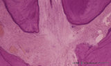 |
Apex of the tooth in osseus socket - alveolus; longitudinal section; human, adult | Stain: Hematoxylin and eosin. From top to bottom: apex region with two tips of dentin covered with cement (darker stained rim; left and right side); note at right tip a dark calcified spot (free cementicle); centrally pulp canal between the tips filled with connective tissue containing blood vessels... | oral cavity; alveolar bone; apical foramen; pulp canal | Poja Histology Collection - Oral Cavity Subset |
| 61 |
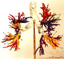 |
Arbor bronchialis, the bronchial tree (human, adult) | Resin corrosion cast of the lower trachea and bronchial tree (posterior aspect). The lobar and segmental bronchi and their main branches are coloured, different colours indicate areas supplied by different segmental bronchi. White-coloured lower trachea (Tr) divides into two principal bronchi (Bp)... | Segmental bronchi ; Macroscopy | Poja Histology Collection - Respiratory System Subset |
| 62 |
 |
Attached denticulus (pulp stone) in the tooth - longitudinal section of root; human, adult. | Stain: Hematoxylin and eosin. From left to right: dentin (purple stained) with dentin tubules; small lightly stained rim of predentin; layers of odontoblasts; irregular structured pulp stone attached to dentin; this false denticle consists of calcified layers with collagen fibers surrounding debris;... | oral cavity; denticulus; pulp stone | Poja Histology Collection - Oral Cavity Subset |
| 63 |
 |
Basal lamina between ameloblasts and odontoblasts in tooth development - advanced bell stage, rat embryo | Stain: Anti-collagen IV immunoperoxidase and hematoxylin counterstained. From top to bottom: Stellate reticulum; Stratum intermedium; Ameloblasts at the distal side oriented towards the basal lamina intermingled with the Korffs' fibers (collagen, brown-black); Odontoblasts; Brown-black stained struc... | oral cavity; basal lamina; collagen IV | Poja Histology Collection - Oral Cavity Subset |
| 64 |
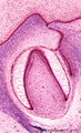 |
Bell stage of the tooth development - human, embryo; low magnification | Stain: Azan. From top to bottom: Stratified ectoderm with a distinct basal layer (red line) of cuboid cells; Dental lamina giving rise to the bell stage (left) and to the primordium of permanent tooth (right); Odontogenic organ (future deciduous tooth) surrounded by fibrous tooth follicle; Out... | oral cavity; dental lamina; predentin | Poja Histology Collection - Oral Cavity Subset |
| 65 |
 |
The bronchial tree and branches of the pulmonary artery (human, adult, posterior aspect) | Resin corrosion cast of left and right lung. Posterior aspect of lower trachea (1, yellow) with two principal bronchi (yellow) with red-stained pulmonary artery (2) and its branching. The cast of both lung lobes reveals especially the intricate divisions and branching of the bronchial tree (yellow) ... | Macroscopy | Poja Histology Collection - Respiratory System Subset |
| 66 |
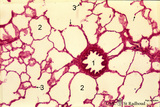 |
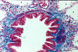
Bronchiolus and alveoli in lung (human, adult) | Stain: Hematoxylin and eosin. (1) indicates a bronchiolus with respiratory epithelium and muscle fibers in its wall. Alveolar spaces (2) are separated by alveolar cell types that lined thin septa with capillaries. The septa end in tips and knobs (3). | Bronchiolus; Alveolar septa; Pneumocyte I; Pneumocyte II; Alveolar cell types | Poja Histology Collection - Respiratory System Subset |
| 67 |
 |
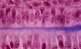
Bronchiolus in the lung (human, adult) | Stain: Azan. The lumen of the bronchiolus is lined with one layer of ciliated epithelium (1) and is folded due to the contraction of smooth muscle fibers (2) in the wall. Note the rich cellularity (3) in the stromal surrounding of the bronchiolus. Alveolar space (4). (5) pulmonary artery branches. | Ciliated epithelium; Bronchiolus | Poja Histology Collection - Respiratory System Subset |
| 68 |
 |
Bronchiolus in the lung (human, adult) | Stain: Azan. The lumen is lined with one layer of ciliated epithelium (1) without goblet cells. Patches of smooth muscle fibers (2) are present. Notice small bronchial arteries (*). Arrows (↓) indicate sites of carbon accumulations between the alveoli (3). | Bronchiolus; Ciliated epithelium | Poja Histology Collection - Respiratory System Subset |
| 69 |
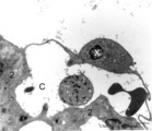 |
Brush cell in lung (rat) | Electron microscopy. In the proximal as well as in the terminal airways a special type of cell the so-called brush(-border) cell could be observed. In this picture this cell (X) is located in the alveolar space. At (*) the brush border, the dense cytoplasm contains many organelles. (C) is a capillar... | Brush cells | Poja Histology Collection - Respiratory System Subset |
| 70 |
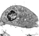 |
Brush cell in lung (rat) | Electron microscopy. In the proximal as well as in the terminal airways a special type of cell can be observed, the so-called brush (-border) cell. In this picture this cell is located in the alveolar space. At (1) the brush border of thick plump microvilli. In the cytoplasm a Gogi area, mitochondri... | Brush cell | Poja Histology Collection - Respiratory System Subset |
| 71 |
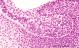 |
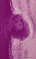
Bud stage outgrowth in tooth development - tooth germ, human, embryo | Stain: hematoxylin. At the top stratified ectoderm continuous with the former epithelial tooth bud that abuts toward the underlying neural crest-derived mesenchymal cells. A basal lamina delimits the palisade-arranged epithelial cells from the surrounding dense aggregate of mesenchymal cells. | oral cavity; tooth bud; tooth development | Poja Histology Collection - Oral Cavity Subset |
| 72 |
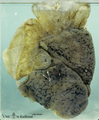 |
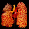
Bullous emphysema of the lung (honeycomb lung, human, adult) | Macroscopy of the left lung showing large apical bullae (3) due to total destruction of all alveoli leaving remnants of the covering pleura. (2) indicates small bullae of foci of destroyed lobules with permanent enlargement of the air spaces and (1) points to spots of anthracosis in less-affected lu... | Macroscopy; Honeycomb lung; Interstitial fibrosis; Hamman-Rich Syndrome | Poja Histology Collection - Respiratory System Subset |
| 73 |
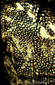 |
Capillary system of a lung alveolus (cat) | Stain: specimen injected with India ink via the pulmonary system. In an en face view of the alveolar wall the black- and granular-stained capillary plexus is well shown. The larger vessels represent branches of the pulmonary arteriole. | Poja Histology Collection - Respiratory System Subset | |
| 74 |
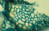 |
Capillary system of an alveolus (cat) | Stain: specimen injected with trypan blue in gelatin via the pulmonary system. In a tangential view the blue-stained dense capillary plexus of one alveolus is well shown. | Poja Histology Collection - Respiratory System Subset | |
| 75 |
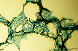 |
Capillary system of lung alveoli (cat) | Stain: specimen injected with trypan blue in gelatin via the pulmonary system. In a tangential view the blue-stained dense capillary plexus surrounding each alveolus is well depicted. | Poja Histology Collection - Respiratory System Subset |