John A. Moran Eye Center Neuro-Ophthalmology Collection: A variety of lectures, videos and images relating to topics in Neuro-Ophthalmology created by faculty at the Moran Eye Center, University of Utah, in Salt Lake City.
NOVEL: https://novel.utah.edu/
TO
| Title | Description | Type | ||
|---|---|---|---|---|
| 51 |
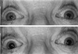 |
Enhanced Mydriasis in Response to Hydroxyamphetamine | Enhanced mydriasis in response to hydroxyamphetamine in a 77-year-old woman with a long-standing, preganglionic, right-sided Horner's syndrome that occurred following cervical neck dissection for thoracic outlet syndrome 30 years earlier. Miosis of the right pupil is apparent in room light (top). Th... | Image |
| 52 |
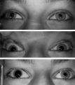 |
Left-sided Horner's Syndrome with an Acquired Preganglionic Localization | Left-sided Horner's syndrome in a 12-year-old girl with an acquired preganglionic localization based on clinical and pharmacologic testing. The cause remained undetermined after extensive radiologic investigations. Left-sided ptosis and miosis are evident in room light (top), and the degree of aniso... | Image |
| 53 |
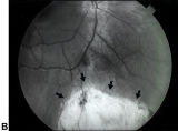 |
Bilateral Iris Colobomas (B) | Bilateral iris colobomas. B. Bilateral colobomatous defects of the inferonasal retina (black arrows) are also present, as shown in the right eye. | Image |
| 54 |
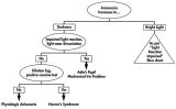 |
Flow Chart for Sorting Out Anisocoria - Bright Light and Darkness | Flow chart for sorting out anisocoria based initially on how it is influenced by bright light and darkness. | Image |
| 55 |
 |
Flow Chart for Sorting Out Anisocoria - Direct Light Reaction of the Pupil | Flow chart for sorting out anisocoria based initially on the integrity of the direct light reaction of the pupil. | Image |
| 56 |
 |
Assessment of an Afferent Pupillary Defect When Only One Iris is Functional | Assessment of an afferent pupillary defect when only one iris is functional. In this example, a right-sided parasellar tumor is compressing both the optic and oculomotor nerves, causing an optic neuropathy and a pupil-involving third crainial nerve palsy. The pupil on the affected side has both an a... | Image |
| 57 |
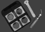 |
Hand-held Equipment Used to Measure a Relative Afferent Pupillary Defect | Hand-held equipment used to measure a relative afferent pupillary defect and to record pupil sizes. Four neutral density filters (0.3, 0.6, 0.9, 1.2 log units) are conveniently carried in a soft cloth carrying pouch. A bright light source (a Finhoff model illuminator is shown here) is ideal for stim... | Image |
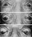
| 58 |
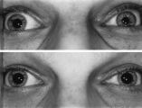 |
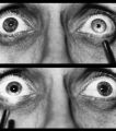
Left-sided Dilation Lag in a Man with Horner's Syndrome | Left-sided dilation lag in a 29-year-old man with Horner's syndrome caused by a posterior mediastinal ganglioneuroma. Note that the degree of anisocoria is greater after 5 seconds in darkness (top) compared with findings after 15 seconds in darkness (bottom). | Image |
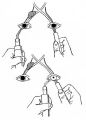
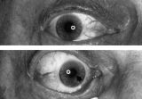
| 59 |
 |
Right-sided Relative Afferent Pupillary Defect | Right-sided relative afferent pupillary defect in a man with optic nerve glioma. When the unaffected left eye is stimulated by light, both pupils constrict (top). When the light is then swung over to the affected right eye, both pupils dilate (bottom). This indicates that pupillomotor conduction thr... | Image |
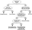
| 60 |
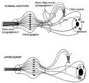 |
Pathophysiology of Signs Associated with a Tonic Pupil | Pathophysiology of signs associated with a tonic pupil. Normally, all parasympathetic fibers of the third cranial nerve synapse in the ciliary ganglion (top). Most postganglionic fibers innervate the ciliary muscle (dashed lines). After injury to the ciliary ganglion, the pupil becomes denervated an... | Image |
| 61 |
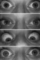 |
Pupil Signs in a 32-year-old Woman with Right-sided Adie's Pupil | Pupil signs in a 32-year-old woman with right-sided Adie's pupil. The right pupil is larger than the left pupil (top), reacts poorly to direct light stimulation (second panel), and better in response to near stimulation (third panel). The right pupil also shows a supersensitive response 30 minutes a... | Image |
| 62 |
 |
Tadpole-shaped Pupil | Tadpole-shaped pupil in a 20-year-old women with frequent episodes of blurred vision and achiness of the right eye lasting several minutes. The patient took a photograph of her eyes during an attack to document the peaked, segmental dilation of her right pupil (black arow). (Thompson HS, Zackon DH, ... | Image |
| 63 |
 |
Argyll Robertson Pupils | Argyll Robertson pupils in an elderly man treated for tabes dorsalis in 1952. His pupils are small and slightly irregular, constrict poorly in response to light stimulation (top), dilate poorly in darkness (middle), but constrict promptly in response to near stimulation (bottom). | Image |
| 64 |
 |
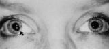
Aberrant Regeneration of the Right Pupil | Aberrant regeneration of the right pupil in a man with a large intracavernous sinus meningioma causing a pupil-involving, incomplete third cranial nerve palsy. His pupil is round when he gazes straight ahead (top). When he tries to rotate the eye medially, the pupil constricts, but a segment of the ... | Image |
| 65 |
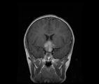 |
See-saw Nystagmus MRI 1 | MRI; See-saw Nystagmus | Image |
| 66 |
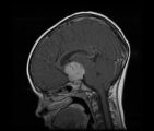 |
See-saw Nystagmus MRI 2 | MRI; See-saw Nystagmus | Image |