John A. Moran Eye Center Neuro-Ophthalmology Collection: A variety of lectures, videos and images relating to topics in Neuro-Ophthalmology created by faculty at the Moran Eye Center, University of Utah, in Salt Lake City.
NOVEL: https://novel.utah.edu/
TO
Filters: Collection: "ehsl_novel_jmec"
| Title | Description | Type | ||
|---|---|---|---|---|
| 51 |
 |
Multifocal Electroretinograms | The most important development in ERGs is the multifocal ERG (mfERG). Erich Sutter adapted the mathematical sequences called binary m-sequences creating a program that can extract hundreds of focal ERGs from a single electrical signal. This system allows assessment of ERG activity in small areas of ... | |
| 52 |
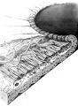 |
Structures of the iris | Structures of the iris. The a indicates the anterior border layer that terminates at the pigmentary ruff of the pupillary border (b). The c indicates the iris sphincter muscle, which is oriented circumferentially within the stroma and located deep to the anterior border layer; d indicates vessels th... | Image |
| 53 |
 |
Dysthyroid Optic Neuropathy: A Preventable Cause of Blindness | Dysthyroid Optic Neuropathy (DON) is a treatable cause of visual loss in ~5% of pts w/ ted. Monitor closely those pts with risk factors (proptosis, tight orbit, restricted motility, strabismus, smoker, diabetic). Oral prednisone is often effective, but frequent relapses after tapering. Orbital xrt ... | |
| 54 |
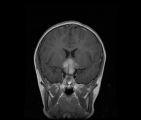 |
See-saw Nystagmus MRI 1 | MRI; See-saw Nystagmus | Image |
| 55 |
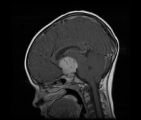 |
See-saw Nystagmus MRI 2 | MRI; See-saw Nystagmus | Image |
| 56 |
 |
Why Don't You See Double? | This presentation was given at the Neurology Grand Rounds in Fall 2011 at the University of Utah. A number of Duane Syndrome cases are covered. Related video can be found in this collection at: Duane's Syndrome Type I: http://content.lib.utah.edu/u?/EHSL-Moran-Neuro-opth,130 Duane's Syndrome Type I... | Text |
| 57 |
 |
The Wall-Eyed Potato Farmer | Young man presenting with apparent episodic neurologic evants that initially was thought to be multiple sclerosis, but as time went on, he had progressive changes in his neurologic exam and in his imaging findings. Brain biopsy revealed Gliomatosis cerebri. Anatomy: Brain Stem; Pons; Midbrain. Patho... | |
| 58 |
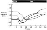 |
Pupillogram Demonstrating Paradoxical Pupillary Constriction to Darkness | Pupillogram demonstrating paradoxical pupillary constriction to darkness in four patients with congenital achromatopsia. Note that the pupils initially constrict when the light is extinguished. (Price MJ, Thompson HS, Judisch GF et al: Pupillary constriction to darkness. Br J Ophthalmol 1981;69:205-... | Image |
| 59 |
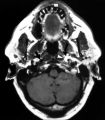 |
Left-sided Internal Carotid Artery Dissection | Left-sided internal carotid artery dissection identified on T-1 weighted magnetic resonance image from a 52-year-old man who suddenly developed left-sided neck and orbital pain along with a droopy left upper eyelid while dragging a deer out of the woods during hunting season. The normal dark flow vo... | Image |
| 60 |
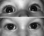 |
Right-sided Pseudo-Horner's Syndrome | Right-sided pseudo-Horner's syndrome in an 8-month-old infant referred because her mother had noted a larger pupil on the left for a few months and her pediatrician thought the right upper lid was droopy. Both pupils reacted normally to light and darkness, the degree of anisocoria was similar in bot... | Image |
| 61 |
 |
Gaze Palsy with Facial Weakness from Pontine AVM | Example of a patient with torsional nystagmus in both eyes and pendular nystagmus in the left eye. Patient is led through instructions for direction of gaze. | Image/MovingImage |
| 62 |
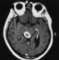 |
An Enhancing Bladder Metastasis Involving the Tectum of the Midbrain | Magnetic resonance image of an enhancing bladder metastasis involving the tectum of the midbrain of a 56-year-old man who developed double vision resulting from skew deviation and divergence insufficiency. He also had a left-sided relative afferent pupillary defect measuring 1.4 log units but showed... | Image |
| 63 |
 |
Ocular Myasthenia | Example of patient with myasthenia gravis. Demonstration of tensilon test. Patient shown to have bilateral ptosis, bilateral duction deficits, and left hypertropia. Discussion of techniques to observe subtle changes, such as bringing in a neutral observer or taking still photographs. Shows split-scr... | Image/MovingImage |
| 64 |
 |
Wall-Eyed Bilateral Internuclear Ophthalmoplegia (WEBINO) | Example of patient with horizontal binocular diplopia. Demonstration of exam, which shows alternating exotropia in cover test. As patient follows object, right eye does not pass the midline as the object moves to the left, while left eye go slightly past the midline, but does not abduct completely. ... | Image/MovingImage |
| 65 |
 |
Wall-Eyed Bilateral Internuclear Ophthalmoplegia (WEBINO) | Example of patient with Wall-Eyed Bilateral Internuclear Ophthalmoplegia. Patient is led through instructions for direction and distance of gaze. | Image/MovingImage |
| 66 |
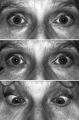 |
Light-near Dissociation | Light-near dissociation in a 51-year-old woman with multiple sclerosis who experienced double vision for 1 week. Her pupils are 5 mm in diameter in room light (top), react poorly in response to direct light reaction (middle), but constrict promptly in response to near stimulation (bottom). She also ... | Image |
| 67 |
 |
Migraine and Cluster Pathophysiology and Treatment | Video lecture covering pathophysiology and treatment of migraine and cluster headaches by Kathleen Digre, MD. | |
| 68 |
 |
The Normal Pupillary Light Reflex | The normal pupillary light reflex is initiated following exposure to light. After a brief latency, both the right (solid line) and left (broken line) pupil constrict, then undergo a small amount of redilation (escape), followed by oscillations (hippus) if the light is sustained. Hippus is not a path... | Image |
| 69 |
 |
Spontaneous Venous Pulsations | This clips shows a spontaneous venous pulsation viewed during an ocular examination. | Image/MovingImage |
| 70 |
 |
Duane's Retraction Syndrome Type 3 | Example of a patient with Type 3 Duane's Retraction Syndrome, as well as bilateral Duane's Syndrome. Shows limitation of abduction in both eyes and adduction in the left eye. Also shows side-view of globe retraction in abduction. | Image/MovingImage |
| 71 |
 |
Introduction to Headache, Migraine and Secondary Headaches | Video lecture covering an introduction to headache, migraine, and secondary headaches by Kathleen Digre, MD. | |
| 72 |
 |
Introduction to the Basic Neurologic Exam | Introduction to the neurological examinations section of NExT. | |
| 73 |
 |
Nutritional Amblyopia | Example of patient with amblyopia with nutritional causes. | Text |
| 74 |
 |
Blepharospasm | Example of patient with blepharospasm. Patient is led through instructions for direction of gaze and opening and closing of eyes. Patient is led through same exercises again after receiving indomethacin treatment. | Image/MovingImage |
| 75 |
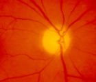 |
4-54a -Optic Neuropathy, Ischemic: Posterior | Image |