The Emory Eye Center Neuro-Ophthalmology Collection contains a variety of lectures, videos and images relating to the discipline of neuro-ophthalmology created by faculty at Emory University in Atlanta, GA.
NOVEL: https://novel.utah.edu/
TO
Filters: Collection: "ehsl_novel_eec"
| Title | Description | Creator | ||
|---|---|---|---|---|
| 51 |
 |
Interpreting Ocular Fundus Photographs: a brief guide | Brief guide for interpreting ocular fundus photographs. | Gabriele Berman, MD; Sachin Kedar, MD; Nancy J. Newman, MD; Valérie Biousse, MD |
| 52 |
 |
Radiologic Appearance of Unilateral Sphenoid Wing Hypoplasia in Neurofibromatosis Type I | MRI features of greater wing sphenoid hypoplasia in the setting of neurofibromatosis type 1. - Figure 1 : Orbital MRI with contrast showing right greater sphenoid wing hypoplasia. The lack of bone tissue leads to herniation of the right temporal lobe into the orbit, pushing forward the orbital conte... | Samuel Bidot, MD; Amit M. Saindane, MD; Valérie Biousse, MD |
| 53 |
 |
Pulsatile Proptosis from Sphenoid Wing Hypoplasia in Neurofibromatosis Type 1 | Clinical and radiologic features of greater wing sphenoid hypoplasia in the setting of neurofibromatosis type 1. Figure 1 : slit lamp examination showing Lisch nodules; Figure 2 : orbit CT scan (1); Figure 3 : orbit CT scan (2) with annotations. For visual examples of this disorder, please see the... | Samuel Bidot, MD; Amit M. Saindane, MD; Valérie Biousse, MD |
| 54 |
 |
Pulsatile Proptosis Profile | See the PowerPoint description for Pulsatile Proptosis From Sphenoid Wing Hypoplasia in Neurofibromatosis Type 1 at: http://content.lib.utah.edu/cdm/ref/collection/ehsl-eec/id/64 See also Pulsatile Proptosis Full Face video: http://content.lib.utah.edu/cdm/ref/collection/ehsl-eec/id/14 | Valérie Biousse, MD |
| 55 |
 |
Direct Carotid-Cavernous Sinus Fistula | A 40-year-old man presented with decreased vision and redness in his left eye. He had a significant trauma to the left side of his face about one year ago, but did not seek medical attention. External examination showed significant proptosis of the left eye (Figure 1) and conjunctival injection and ... | Jonathan A. Micieli, MD; Valérie Biousse, MD |
| 56 |
 |
Sturge-Weber Syndrome | A case of Sturge-Weber syndrome (Encephalotrigeminal angiomatosis) with angiomas that involve the leptomeninges, and the skin of the ipsilateral hemiface, associated with congenital glaucoma in the same eye. Various illustrations are included to demonstrate the port wine stain, enlarged optic nerve ... | Supharat Jariyakosol, MD; Valérie Biousse, MD |
| 57 |
 |
Ocular Hypoperfusion from Severe Internal Carotid Artery Stenosis | 68 year-old man complaining of mildly decreased vision OD with fluctuation of vision throughout the day. Fluorescein angiography shows delayed choroidal and retinal fillings, suggesting hypoperfusion of the right eye. | Samuel Bidot, MD; Amit M. Saindane, MD; Valérie Biousse, MD |
| 58 |
 |
Occipital Infarction with Incomplete Congruent Homonymous Hemianopia | CT appearance of a remote occipital infarction. Congruent homonymous hemianopia. | Samuel Bidot, MD; Amit M. Saindane, MD; Valérie Biousse, MD |
| 59 |
 |
Terson Syndrome and Subarachnoid Hemorrhage | A case of Terson syndrome resulting with subarachnoid hemorrhage and right vitreous hemorrhage resulting from a left pericallosal artery aneurysm. Figure 1 : External photograph of right eye demonstrates blunted red reflex secondary to vitreous hemorrhage Figure 2 : External photograph of left eye d... | Joshua Levinson, MD; Valérie Biousse, MD |
| 60 |
 |
Terson Syndrome With Cranial Nerve 3 Palsy Due to Subarachnoid Hemorrhage from Arteriovenous Malformation and Aneurysmal Rupture | A case of Terson syndrome due to AVM and posteral cerebral aneurysm. The patient developed a left CN3 palsy due to hematoma involving the left midbrain. Figure 1 : External photograph of right eye demonstrates blunted red reflex secondary to vitreous hemorrhage Figure 2 : External photograph of lef... | Joshua Levinson, MD; Valérie Biousse, MD |
| 61 |
 |
Internal Carotid Artery / Posterior Communicating Artery Aneurysm with Third Nerve Palsy Shown on CT Angiogram | Internal Carotid Artery / Posterior Communicating Artery Aneurysm with Third Nerve Palsy Shown on CT Angiogram ; anatomic description of vascular and bony findings on the CTA. - Figure 1 : 51 year-old man complaining of painful binocular diplopia. Orange arrows indicate the direction of gaze. In p... | Samuel Bidot, MD; Amit M. Saindane, MD; Valérie Biousse, MD |
| 62 |
 |
Sellar Aneurysm with Chiasmal Compression | This is a case of aneurysm of the internal carotid artery, invading the sella and complicated by chiasmal compression and bitemporal hemianopia. Figure 1 : Humphrey visual fields (gray scale and pattern deviations) Figure 2a : T1-weighted axial brain MRI (1): well defined circular intracerebral mass... | Rabih Hage, MD; Valérie Biousse, MD |
| 63 |
 |
Large Right Hypophyseal Aneurysm Causing a Junctional Scotoma | Right, multi-lobulated superior hypophyseal artery aneurysm measuring 1.6 x 1.2 x 2.2 cm with 6 mm neck causing a right junctional scotoma . Images from a brain CT with contrast, a brain CT angiography with contrast, cerebral angiogram, Humphrey visual fields and ocular fundus photographs are includ... | Laurel N. Vuong, MD; Valérie Biousse, MD |
| 64 |
 |
Previous Branch Retinal Artery Occlusion | This is a typical case of an old branch retinal artery occlusion in a 64 year old woman presenting with persistent monocular vision loss. She had sudden onset of painless vision loss in the inferior field of her left eye approximately one year prior. Her past medical history was significant for atri... | Benson S. Chen, MBChB FRACP; Valérie Biousse, MD |
| 65 |
 |
Choroidal Infarction in Giant Cell Arteritis | An 80-year-old Caucasian woman presented with a 10-day history of headaches, intermittent binocular diplopia, and jaw pain. Temporal artery biopsy confirmed a diagnosis of giant cell arteritis. Examination showed characteristic large area of choroidal ischemia that is well-known to be associated wit... | Wael A. Alsakran, MD; Andre Aung, MD; Valérie Biousse, MD |
| 66 |
 |
Cotton Wool Spots in Giant Cell Arteritis | This is a case of cotton wool spots in a patient with temporal artery-biopsy proven temporal arteritis.; ; A 66-year-old woman presents with isolated painless vision loss related to a left optic neuropathy in her left eye. She denies systemic symptoms to suggest giant cell arteritis.; Her examinatio... | Rahul A. Sharma, MD, MPH; Valérie Biousse, MD |
| 67 |
 |
Classic Pathology Findings in Giant Cell Arteritis | An 80-year-old Caucasian woman presented with a 10 day history of headaches, intermittent binocular diplopia, and jaw pain. Temporal artery biopsy confirmed a diagnosis of giant cell arteritis. Pathology findings were classic for giant cell arteritis with numerous inflammatory cells in the tunica me... | Andre Aung, MD; Corrina Azarcon, MD; Wael A. Alsakran, MD; Valérie Biousse, MD |
| 68 |
 |
MRI Findings in Giant Cell Arteritis | Case 1. An 80-year-old Caucasian woman presented with a 10-day history of headaches, intermittent binocular diplopia, and jaw pain. Temporal artery biopsy confirmed a diagnosis of giant cell arteritis. MRI with contrast showed enhancement of bilateral optic nerve sheaths in addition to enhancement o... | Wael A. Alsakran, MD; Andre Aung, MD; Valérie Biousse, MD |
| 69 |
 |
Occipital Pyogenic Abscess with Homonymous Hemianopia | This is a case of right occipital abscess with a left homonymous hemianopia. Number of Figures and legend for each: 8 figures Figure 1: Humphrey visual fields: Dense left homonymous hemianopia Figure 2: T2-weighted axial MRI : Round, hyperintense lesion (yellow arrow) in the right occipital lobe sur... | Rabih Hage, MD; Valérie Biousse, MD |
| 70 |
 |
Ophthalmic Artery Aneurysm | Slideshow describing ophthalmic artery aneurysm with MRI imaging. | Valérie Biousse, MD |
| 71 |
 |
Funduscopic Findings of Acute Central Retinal Artery Occlusion | A 59-year-old man was referred for assessment acute vision loss in the right eye. His examination showed: best-corrected visual acuity: light perception OD, 20/20 OS; pupils: Relative afferent pupillary defect OD; color vision: unable to visualize control plate OD, 14/14 OS correct Ishihara plates. ... | David B. Enfield, MD; Valérie Biousse, MD |
| 72 |
 |
Neurosarcoidosis | This is an illustrated guide to the clinical diagnosis of neurosarcoidosis. Sarcoidosis is a chronic systemic inflammatory disorder characterized by non-caseating granulomas. It can affect multiple organ systems including the lungs, skin, orbit, and brain. When there is central nervous system (CNS) ... | Bryce Buchowicz, MD; Valérie Biousse, MD |
| 73 |
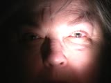 |
Left RAPD | Video clip displaying pupillary examination and RAPD measurement. | Valérie Biousse, MD |
| 74 |
 |
Bilateral Lens Subluxation in Marfan Syndrome | This is a case of known Marfan syndrome with bilateral progressive visual loss. The ocular examination showed bilateral lens dislocation. Figure 1a: Typical superonasal lens subluxation in both eyes Figure 1b: The arrows show the inferior edges of the lenses Figure 2: Optical section of the lenses u... | Rabih Hage, MD; Valérie Biousse, MD |
| 75 |
 |
Iris Transillumination | Single case of iris transillumination in a patient with albinism. Figure 1 : Anterior segment photograph demonstrating reddish hue to iris in albinism Figure 2 : Slit lamp photograph with retroillumination demonstrating iris transillumination Figure 3 : Slit lamp photograph with retroillumination... | Jason Peragallo, MD; Valérie Biousse, MD |
