The Health Education Assets Library (HEAL) is a collection of over 22,000 freely available digital materials for health sciences education. The collection is now housed at the University of Utah J. Willard Marriott Digital Library.
TO
Filters: Collection: "ehsl_heal"
| Title | Description | Subject | Collection | ||
|---|---|---|---|---|---|
| 51 |
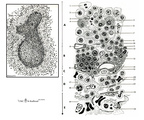 |
Scheme electron microscopy of the border of secondary nodule/red pulp in spleen | The left figure POJA-L974 shows a scheme of histological impression of a survey of a secondary splenic follicle or nodule. The rectangle is enlarged in the right figure POJA-L976B and shows the following elements: A. germinal centre; B. mantle zone; C. dendritic cell area; D. marginal zone; E. ... | white pulp; marginal zone; red pulp; antigen presenting cell (APC) | Poja Histology Collection - Lymphatic Tissues and Organs Subset |
| 52 |
 |
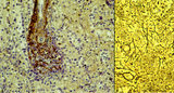
The effect of cyclophosphamide on the CD8-thymocytes in thymus (rat) | Stain: Immunoperoxidase staining with diaminobenzidin (DAB) and hematoxylin counterstained on frozen section. A single injection with cyclophosphamide (CP, 70 mg/ml) induces a transient cortical involution after 4 days, i.e. the darkly stained cortex and the lightly stained medulla in normal thymus ... | cyclophosphamide; CD8 monoclonal antibody; immunosuppression; lymphoid tissue | Poja Histology Collection - Lymphatic Tissues and Organs Subset |
| 53 |
 |
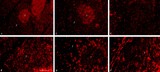
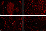
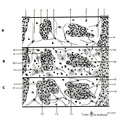
Detailed localization of heparan sulfate (HS) in spleen (rat) | Stain: Immunofluorescence of Alexa 594 red labeled single chain antibody 3C3 for heparan sulfate (HS). The antibody stains HS epitopes of the meshwork of reticulum cells and the basal membrane of blood vessels. A: survey white pulp spleen. B: marginal sinuses between PALS area and red pulp. C: red... | white pulp; PALS; heparan sulfate | Poja Histology Collection - Lymphatic Tissues and Organs Subset |
| 54 |
 |
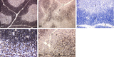
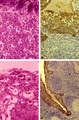
Immunohistochemistry with cellular markers in thymus (rat) | Stain: Alexa-594 red immunofluorescence. (1) medulla. (2) cortex. (A-survey, B-cortex): ER13 antibody stains for MHC-class II antigens on reticular cell types in medulla and cortex. (C): ED1 monoclonal antibody stains a single chain glycoprotein of 110 kDa on the lysosomal membrane of myeloid cel... | ER13 antibody; ED1 monoclonal antibody; ED2 monoclonal antibody; ER2 monoclonal antibody | Poja Histology Collection - Lymphatic Tissues and Organs Subset |
| 55 |
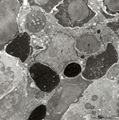 |
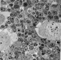
Phagocytosis in splenic red pulp (mouse) | Electron microscopy. Stain: Peroxidase reaction with diaminobenzidin staining. A diversity of red blood cells in the red pulp can be discerned due to the DAB staining of hemoglobin by oxidized benzidin (dark and light staining). The macrophage (1) shows peroxidase activity along the nuclear membran... | electron microscopy; phagocytosis | Poja Histology Collection - Lymphatic Tissues and Organs Subset |
| 56 |
 |
Red pulp of spleen with venous sinusoids (monkey, human) | Stain: A: Silver stain (Movat) (monkey); B: Silver stain (Gomori) (human). (A): The darkly stained fibres are conspicuous in the PALS (1) area arranged in parallel rows. The blood vessels continue in the surrounding splenic sinusoids (4). The wall of the sinusoid is built as a grid, the space is su... | sinusoid; reticular fibres | Poja Histology Collection - Lymphatic Tissues and Organs Subset |
| 57 |
 |
Survey of pharyngeal tonsil ('lymphoepithelial tissues', 'gut-associated lymphatic tissue' or GALT) (human) | Stain Azan. The solitary pharyngeal tonsil is localized in the pharyngeal fornix and belongs to the so-called Waldeyer's ring of pharyngeal lymphatic tissue. A survey shows the folded columnar epithelium (1) with faintly light-stained goblet cells with clefts (2) in between. The lamina propria co... | pharyngeal tonsil; GALT | Poja Histology Collection - Lymphatic Tissues and Organs Subset |
| 58 |
 |
Cystic corpuscle in thymus (human, adult) | Stain: Azan. With age the remaining Hassall's corpuscles become more prominent and might represent large multiform structures of over hundreds of microns. The central cells (*) are keratinized. They might be swollen, calcify and become necrotic eventually, or undergo lysis, leaving a large cystic st... | thymic corpuscle (Hassalls); epithelioreticular cell (ERC); lymphoid tissue | Poja Histology Collection - Lymphatic Tissues and Organs Subset |
| 59 |
 |
Immunohistochemistry of thymus (rat) | Stain: Alexa-594 red immunofluorescence. (1) Medulla. (2) Cortex. (A): Staining with a monoclonal antibody against vimentin illustrates that vimentin is localised in the stromal cells of the connective tissue of the capsule, of the septa or trabeculae that invade the cortex (2) and overwhelmingly ... | vimentin; laminin; fibronectin; ED2 monoclonal antibody | Poja Histology Collection - Lymphatic Tissues and Organs Subset |
| 60 |
 |
Lymph node (human) | Stain: Azan. Left and right: survey of lymph nodes, compare the individual differences between the nodes. The pictures display the cortex (1) with capsule (3), and the medulla (2). The cortex consists of (secondary) follicles displaying a clear germinal centre (4). The paracortex (T cell area) is ... | follicle; paracortex; cortex; germinal center | Poja Histology Collection - Lymphatic Tissues and Organs Subset |
| 61 |
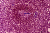 |
Spleen with central artery in secondary lymphatic nodule (human) | Stain: Azan. The white pulp consists of (1) a cross-section of the central artery, (2) the germinal centre with few branches of the central artery; the mantle zone (3) and the marginal zone (4) surrounded by a perilymphoid zone around the nodule. The perilymphoid zone is composed of concentricall... | germinal center; PALS; central artery; marginal zone | Poja Histology Collection - Lymphatic Tissues and Organs Subset |
| 62 |
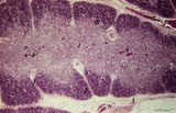 |
Thymus (human, newborn) | Stain: Hematoxylin & eosin. The thymus is a bilobed lymphoepithelial organ derived as an outgrowth from the third branchial (pharyngeal) pouch, and situated in the anterior mediastinum. Each lobe is divided into multiple lobules by fibrous septa or trabeculae (3). Each lobule consists of an outer co... | epithelioreticular cells (ERC); thymus hormones; Hassalls corpuscle ; lymphoid tissue | Poja Histology Collection - Lymphatic Tissues and Organs Subset |
| 63 |
 |
The effect of cyclophosphamide on CD3-thymocytes in thymus (rat) | Stain: Immunoperoxidase staining with diaminobenzidin (DAB) and hematoxylin counterstained on frozen section.. A single injection with cyclophosphamide (CP, 70 mg/ml) induces a transient cortical involution after 4 days, i.e. the dark-blue stained cortex and the lightly stained medulla in normal thy... | cyclophosphamide; CD3 monoclonal antibody; lymphoid tissue ; immunosuppression | Poja Histology Collection - Lymphatic Tissues and Organs Subset |
| 64 |
 |
Follow-up of the process of diapedesis through stratified epithelium of the palatine tonsil (gut-associated lymphatic tissue or GALT) (human) | Stain: (A, C) Azan and (B, D) anti-keratin-antibody and immunoperoxidase staining using diaminobenzidin (DAB) on frozen section, counterstained with hematoxylin. (1) (keratinized) stratified squamous epithelial cells. (2) between desquamated epithelial cells the spaces are filled with infiltrated l... | stratified squamous epithelial cells; GALT | Poja Histology Collection - Lymphatic Tissues and Organs Subset |
| 65 |
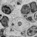 |
Thymus cortex (rat, young adult) | Electron microscopy. Within the thymic cortex a type II epithelioreticular cell or so-called (sometimes multinucleated) thymic nurse cell (TNC) shows a characteristic electron-light nucleus and nucleolus (1). The branches are squeezed between the thymocytes (2). In the cytoplasm a variety of empty a... | MHC class I and II expression; epithelioreticular cell type I and II; thymic nurse cell TNC; lymphoid tissue | Poja Histology Collection - Lymphatic Tissues and Organs Subset |
| 66 |
 |
Corpuscles in thymus (human, puberty) | Stain: Hematoxylin. A: Several thymic (Hassall's) corpuscles (*) of varying sizes within the medulla (1). The Hassall bodies are surrounded by recognizable flattened cells showing keratohyalin granules. The outer shell consists of more layers of close packed concentrically arranged cells with light-... | thymic corpuscle (Hassalls); epithelioreticular cell (ERC); thymus medulla; lymphoid tissue | Poja Histology Collection - Lymphatic Tissues and Organs Subset |
| 67 |
 |
Scheme of the thymus during ageing (human) | Physiological changes in the course of human ageing lead to almost complete degeneration of the thymus (age involution). Both cortex and medulla become depleted of lymphocytes, and the distinction between both layers gradually becomes less. The remaining reticular meshwork of connective tissue is gr... | thymus involution; thymus development; thymus age; lymphoid tissue | Poja Histology Collection - Lymphatic Tissues and Organs Subset |
| 68 |
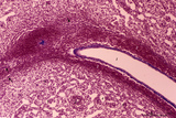 |
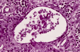
Spleen with central artery in lymphatic nodule (human) | Stain: Azan. A longitudinally cut central artery (1) of a lymphatic nodule or follicle, (white pulp) is invested by a distinct lymphatic sheath (PALS) composed of concentric layered T lymphocytes) (2). The red pulp consists of open venous sinusoids (4) and splenic cords (5, Billroth) with macrophage... | central artery; PALS; sinusoid ; Billroth | Poja Histology Collection - Lymphatic Tissues and Organs Subset |
| 69 |
 |
Survey of spleen (human) | Stain: Trichrome (Goldner). Encapsulated by a relatively thin fibroelastic capsule (1), the splenic parenchyma shows a preferable peripheral location of the red pulp (3). There are many secondary lymphatic nodules (2) as part of the white pulp, all showing a clear germinal centre. The red pulp is a ... | white pulp; red pulp | Poja Histology Collection - Lymphatic Tissues and Organs Subset |
| 70 |
 |
Survey of the border of splenic white and red pulp (rat) | Electron microscopy. Megakaryocytes (1) are commonly found in adult spleen of rodents. In this area they are located just at the border of the white pulp (WP) with a variety of lymphocytes (3). In the splenic cords (Billroth) of the red pulp (RP) erythrocytes (2) are found. | electron microscopy; white pulp; Billroth | Poja Histology Collection - Lymphatic Tissues and Organs Subset |
| 71 |
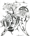 |
Scheme of blood circulation in the red pulp in the spleen | A. closed circulation B. open circulation A. The route of closed circulation proposes that blood empties from the capillaries directly into the splenic sinus. The central artery bifurcates into penicillar arterioles (1) and the blood slowly enters ensheathed capillaries, surrounded by agg... | white pulp; marginal zone; red pulp; open circulation | Poja Histology Collection - Lymphatic Tissues and Organs Subset |
| 72 |
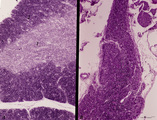 |
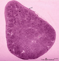
Accidental involution of thymus (mouse, malaria infection) | Stain: Hematoxylin & eosin. Due to the infection with malarial parasites (Plasmodium berghei in mice) a steroid-related involution of the thymus is induced in mice within 14 days. A: normal thymus with cortex (2) and medulla (1). B: There is still a quite large remnant of the original thymus tissue ... | malaria infection; thymus involution; Plasmodium berghei; lymphoid tissue | Poja Histology Collection - Lymphatic Tissues and Organs Subset |
| 73 |
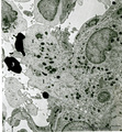 |
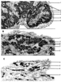
Border of a marginal zone in spleen (rat) | Stain: Immunoelectron microscopy (gold labeling of heparan sulfate in Lowicryl embedding, using the single chain antibody HS4C3). The zone of red pulp immediately surrounding a lymphatic nodule is called the marginal zone (perilymphoid zone) and is composed of a scaffold of basal lamina material w... | marginal zone; immuno electron microscopy; heparan sulfate; dendritic cell | Poja Histology Collection - Lymphatic Tissues and Organs Subset |
| 74 |
 |
Scheme of the medullar changes in lymph node after antigen stimulation | Upon antigenic stimulation the reticular cells that line the medullar sinusoids start to phagocytise the antigens (i.e. infective agents) and change from small tiny stellate cells into swollen large cells due to phagocytosis. They detach from the medullar sinus wall. After ten days the situation has... | follicle; antigen stimulation; scheme; germinal center | Poja Histology Collection - Lymphatic Tissues and Organs Subset |
| 75 |
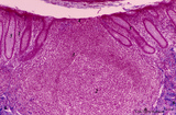 |
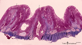
Appendix ('gut-associated lymphatic tissue' or GALT) (human) | A large nodule in the appendix extends through the proper lamina (1) and submucosa. The nodule is similar to that found in a lymph node with germinal centre (2) and darker-stained cap (crescent) (3) orientated towards the lumen of the gut showing a flattened dome area covered with discontinuous epit... | GALT; follicle; lymphoid tissue | Poja Histology Collection - Lymphatic Tissues and Organs Subset |