John A. Moran Eye Center Neuro-Ophthalmology Collection: A variety of lectures, videos and images relating to topics in Neuro-Ophthalmology created by faculty at the Moran Eye Center, University of Utah, in Salt Lake City.
NOVEL: https://novel.utah.edu/
TO
| Title | Description | Type | ||
|---|---|---|---|---|
| 51 |
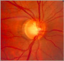 |
Notching of the Neuro-retinal Rim | The neuro-retinal rim becomes thinner; in particular the rim superotemporally and inferortemporally may develop a notch which is usually superior or inferior and rarely nasal or temporal. These notches are believed to be due to focal ischemic damage to the neuro-retinal rim. Glaucoma with Notching a... | Image |
| 52 |
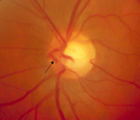 |
3-65 - Shunt Vessels (Glaucoma) | Chronic end-stage glaucoma produces high pressure that interferes with venous drainage from the disc and broad smooth venous collaterals drain the disc centrifugally to the disc margin where they drain. | Image |
| 53 |
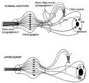 |
Pathophysiology of Signs Associated with a Tonic Pupil | Pathophysiology of signs associated with a tonic pupil. Normally, all parasympathetic fibers of the third cranial nerve synapse in the ciliary ganglion (top). Most postganglionic fibers innervate the ciliary muscle (dashed lines). After injury to the ciliary ganglion, the pupil becomes denervated an... | Image |
| 54 |
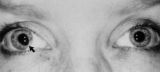 |
Tadpole-shaped Pupil | Tadpole-shaped pupil in a 20-year-old women with frequent episodes of blurred vision and achiness of the right eye lasting several minutes. The patient took a photograph of her eyes during an attack to document the peaked, segmental dilation of her right pupil (black arow). (Thompson HS, Zackon DH, ... | Image |
| 55 |
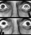 |
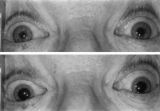
Right-sided Relative Afferent Pupillary Defect | Right-sided relative afferent pupillary defect in a man with optic nerve glioma. When the unaffected left eye is stimulated by light, both pupils constrict (top). When the light is then swung over to the affected right eye, both pupils dilate (bottom). This indicates that pupillomotor conduction thr... | Image |
| 56 |
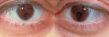 |
Bilateral Iris Colobomas | Coloboma literally means a "gap"-and can be used to describe any fissure, hole, or gap in the eye. The term most often is used to refer to a congenital gap in the disc, retina, the choroid, and the iris. Colobomas occur because the embryonic fissure fails to fuse. Since the fissure closure begins in... | Image |
| 57 |
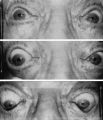 |
Argyll Robertson Pupils | Argyll Robertson pupils in an elderly man treated for tabes dorsalis in 1952. His pupils are small and slightly irregular, constrict poorly in response to light stimulation (top), dilate poorly in darkness (middle), but constrict promptly in response to near stimulation (bottom). | Image |
| 58 |
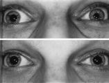 |
Left-sided Dilation Lag in a Man with Horner's Syndrome | Left-sided dilation lag in a 29-year-old man with Horner's syndrome caused by a posterior mediastinal ganglioneuroma. Note that the degree of anisocoria is greater after 5 seconds in darkness (top) compared with findings after 15 seconds in darkness (bottom). | Image |
| 59 |
 |
Enhanced Mydriasis in Response to Hydroxyamphetamine | Enhanced mydriasis in response to hydroxyamphetamine in a 77-year-old woman with a long-standing, preganglionic, right-sided Horner's syndrome that occurred following cervical neck dissection for thoracic outlet syndrome 30 years earlier. Miosis of the right pupil is apparent in room light (top). Th... | Image |
| 60 |
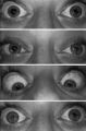 |
Pupil Signs in a 32-year-old Woman with Right-sided Adie's Pupil | Pupil signs in a 32-year-old woman with right-sided Adie's pupil. The right pupil is larger than the left pupil (top), reacts poorly to direct light stimulation (second panel), and better in response to near stimulation (third panel). The right pupil also shows a supersensitive response 30 minutes a... | Image |
| 61 |
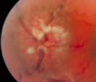 |
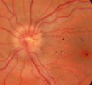
3-35a - Papilledema Stages | Grading Papilledema: Stage 4 Stage 4 = Complete obliteration of the cup and complete obscuration of at least some vessels on the surface of the disc. There may be small dilated capillaries on the disc that resemble telangiectasia. It is not the NFL infarcts or hemorrhages but the obscuration of the ... | Image |
| 62 |
 |
3-31b - Papilledema Stages | Grading Papilledema: Stage 0 GRADING PAPILLEDEMA GRADING PAPILLEDEMA We grade papilledema in order to tell us how severe it is. The most sensible grading scheme has been provided by Lars Frisén. STAGE 0: This woman had documented increased intracranial pressure of 340 mm water. Very little if any ... | Image |
| 63 |
 |
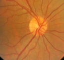
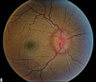
3-32b - Papilledema Stages | Grading Papilledema: Stage 1 Stage 1 = C shaped blurring of the nasal, superior and inferior borders. Usually the temporal margin is normal. Also notice the chorio-retinal folds (arrows) that eminate toward the macula (m) | Image |
| 64 |
 |
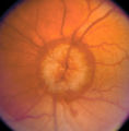
3-34c Papilledema Stages | Grading Papilledema: Stage 3 Stage 3 = Elevation of the entire disc with partial obscuration of the retinal vessels at the disc margin. Here the vessels are partly obscured and make the development into stage 3 easier to call. | Image |
| 65 |
 |
Stage 2 - Papilledema | Image | |
| 66 |
 |
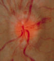
3-36a - Papilledema Stages | Grading Papilledema: Stage 5 Stage 5 = Dome-shaped appearance with all vessels being obscured. (Sometimes called "champagne cork" swelling--because of its dome shape.) | Image |