The Health Education Assets Library (HEAL) is a collection of over 22,000 freely available digital materials for health sciences education. The collection is now housed at the University of Utah J. Willard Marriott Digital Library.
TO
Filters: Collection: ehsl_heal
| Title | Description | Subject | Collection | ||
|---|---|---|---|---|---|
| 626 |
 |
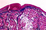
Uvula (soft palate, human; low magnification) | Stain: Azan. At the right (oral side, non-keratinized stratified squamous epithelium); at the left (nasal side, same epithelium). In the submucosa mainly mucous uvular glands are localized. At the bottom, striated muscle of the uvular muscle. The uvular tip is richly vascularized. | oral cavity | Poja Histology Collection - Oral Cavity Subset |
| 627 |
 |
Uvula (soft palate, human) | Stain: Azan. Detail of the uvula at the nasal side shows the border between the non-keratinized stratified squamous epithelium (right, oral side) and pseudostratified columnar epithelium (left, nasal side). The lamina propria is composed of dense connective tissue. At the bottom part of a blood-fil... | oral cavity; lining mucosa | Poja Histology Collection - Oral Cavity Subset |
| 628 |
 |
Uvula (soft palate, human) | Stain: Azan. Detail of the uvula at the oral side; non-keratinized stratified squamous epithelium. Note richly vascularized lamina propria. In the submucosa mainly mucous uvular glands with draining ducts. | oral cavity; lining mucosa; mucous glands | Poja Histology Collection - Oral Cavity Subset |
| 629 |
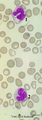 |
Vacuolar degeneration of neutrophils in peripheral blood smear (human) | Stain: May-Grnwald-Giemsa (MGG). Toxic degeneration of neutrophil resulting in vacuolization of cytoplasm (1); cell with beginning vacuolization (2). | Poja Histology Collection - Blood & Bone Marrow Subset | |
| 630 |
 |
Venous circulation pattern in perfused spleen (human) | Stain: Azan. The composed picture shows part of the splenic circulation system at several enlargements (inset, A, B). The open venous sinusoids (1) drain via short pulp veins into thin-walled trabecular veins (2), subsequently into thick-walled trabecular veins (4). The trabeculae originate from the... | splenic circulation; trabecular veins; sinusoid ; PALS | Poja Histology Collection - Lymphatic Tissues and Organs Subset |
| 631 |
 |
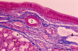
Ventricular fold (plica ventricularis) (human) | Stain: Azan. On top pseudostratified epithelium (1). Lamina submucosa with predominantly mucous glands (3), fat cells (4) and blood vessels. The lamina propria also contains lymphoid follicles (2) with a germinal center (immune protection). | Laryngeal glands; Ventricular fold; Seromucous glands | Poja Histology Collection - Respiratory System Subset |
| 632 |
 |
Ventricular fold (plica ventricularis) (human, higher magnification) | Stain: Azan. On top pseudostratified columnar, ciliated epithelium (1). Centrally below an excretory duct (2) of laryngeal seromucous gland (3) with some adipocytes (*). Blood vessels and two lymph vessels (4) are present. | Supraglottis; Ventricular fold; Laryngeal glands; Seromucous glands | Poja Histology Collection - Respiratory System Subset |
| 633 |
 |
Villi of complete hydatidiform mole (human) | (A) Macroscopy aggregation (1) of abnormal villi of hydatidiform mole intermingled with blood clots (2). (B) After dissection of a complete mole grape-like aggregations of dilated chorionic villi are presented as numerous liquid-filled vesicles (1) varying in diameters (from few mm up to 1 cm) surr... | placenta; trophoblast; hydatiform mole | Poja Histology Collection - Placenta |
| 634 |
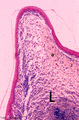 |
Vocal cord (plica vocalis) (human, glottis) | Stain: Azan. The true vocal cord consist of the vocal ligament (L) covered by stratified squamous epithelium and the striated vocal muscle (medial part of the thyreoarythenoid muscle, not depicted in this picture). At the tip of the vocal cord the stratified epithelium is distinct and the lamina pro... | Vocal ligament | Poja Histology Collection - Respiratory System Subset |
| 635 |
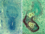 |
White pulp of spleen (mouse) | Stain: Hematoxylin & eosin in A and alkaline phosphatase in B (with substrate Naphtol Fast Blue RR). The general structure of the white pulp of the spleen and its specific microenvironment for T and B cells is well illustrated using alkaline phosphatase that strongly stains the capillaries around th... | alkaline phosphatase; PALS; T lymphocytes; white pulp | Poja Histology Collection - Lymphatic Tissues and Organs Subset |