The Health Education Assets Library (HEAL) is a collection of over 22,000 freely available digital materials for health sciences education. The collection is now housed at the University of Utah J. Willard Marriott Digital Library.
TO
Filters: Collection: ehsl_heal
| Title | Description | Subject | Collection | ||
|---|---|---|---|---|---|
| 601 |
 |
Thymus medulla (rat, neonate) | Electron microscopy. An interdigitating cell in the thymic corticomedullary region shows a large electron-light cytoplasm with a complex branching (7) at the periphery. The nucleus is sectioned twice (1). There is abundance of organelles as well as of quite uniform electron-dense lysosomal structure... | medullar epithelioreticular cell; interdigitating cell; corticomedullar region; lymphoid tissue | Poja Histology Collection - Lymphatic Tissues and Organs Subset |
| 602 |
 |
Thymus medulla (rat, young adult) | Electron microscopy. Epithelioreticular cells of the medulla (1) close to each other. The electron-light cytoplasm contains many small vesicles (1, Golgi area) as well as cross-sections of vacuoles (2) with small finger-like cytoplasmic extrusions in the lumen. Electron-dense lysosomal structures (3... | medullar epithelioreticular cell ; thymus medulla; lymphoid tissue | Poja Histology Collection - Lymphatic Tissues and Organs Subset |
| 603 |
 |
Thymus medulla (rat, young adult) | Electron microscopy. Surrounded by thymocytes (3) a medullary macrophage with an electron-light nucleus (1). The cytoplasm contains many electron-dense lysosomes of varying sizes and forms (2). | medullar macrophage; lymphoid tissue | Poja Histology Collection - Lymphatic Tissues and Organs Subset |
| 604 |
 |
Thymus medulla (rat, young adult) | Electron microscopy. Epithelioreticular cell of the medulla with an electron-light cytoplasm contains cross-sections of vacuoles with small finger-like cytoplasmic extrusions in the lumen (1). Electron-dense lysosomal structures (2) are also present as well as bundles of intermediate filaments (kera... | medullar epithelioreticular cell ; keratin filaments; lymphoid tissue | Poja Histology Collection - Lymphatic Tissues and Organs Subset |
| 605 |
 |
Tongue (lingual gland, human) | Stain: Azan. The posterior lingual gland is composed of areas with pure serous acini neighbouring pure mucous acini between the tongue muscles (at the left few striated fibers). | oral cavity; lingual gland | Poja Histology Collection - Oral Cavity Subset |
| 606 |
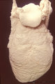 |
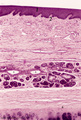
Tongue (macroscopy, dorsal side, human) | At the bottom (apex) of the picture the dorsal side is covered with numerous closed packed, small, conical filiform papillae. Interspersed among them are the larger fungiform papillae. Posteriorily the circumvallatae papillae are arranged in a V-shape row. Behind the V-row the area is covered with l... | oral cavity; papillae | Poja Histology Collection - Oral Cavity Subset |
| 607 |
 |
Tongue (ventral part, human) | Stain: Hematoxylin and eosin. Non-keratinized squamous epithelium, followed by a small lamina propria and the intrinsic striated lingual muscles. | oral cavity; lingual muscles | Poja Histology Collection - Oral Cavity Subset |
| 608 |
 |
Tongue Papillae Scheme - tongue, human, adult | A. Scheme survey dorsal surface of tongue (human, adult). Magnification 35x. B. Scheme of filiform and fungiform papillae (tongue, human, adult). Magnification 35x. 1. foliate papillae (rudimentary in human); 2. circumvallate papillae; 3. foramen cecum; 4. palatine tonsils; 5. filiform ... | oral cavity; papillae | Poja Histology Collection - Oral Cavity Subset |
| 609 |
 |
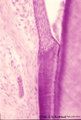
Tooth ('free' gingiva of decalcified tooth, human, adult) | Stain: Hematoxylin and eosin. Oral side (left) with stratified squamous epithelium, at the right side part of a tooth (dentin), at the bottom side part of the alveolar bone. The epithelium shows slight parakeratosis with many narrow deep papillae of the lamina propria (oral side). On the tooth-relat... | oral cavity; junctional epithelium; alveolar bone; gingival sulcus | Poja Histology Collection - Oral Cavity Subset |
| 610 |
 |
Tooth ('free' gingiva of decalcified tooth, human, adult) | Stain: Hematoxylin and eosin. On top part of the gingival sulcus, at the left stratified junctional epithelium normally attached to the surface of the enamel (dissolved due to decalcification). Top left shows foci of chronic inflammatory cell infiltration, left side faint stained bundle of fibers (p... | oral cavity; acellular cementum; junctional epithelium | Poja Histology Collection - Oral Cavity Subset |
| 611 |
 |
Tooth ('free' gingiva of decalcified tooth, human, adult) | Stain: Hematoxylin and eosin. On top the gingival sulcus; at the left stratified junctional epithelium normally attached to the surface of the enamel (dissolved due to decalcification). Below the epithelium a focus of chronic inflammatory cell infiltration, followed by a bundle of fibers of the per... | oral cavity; acellular cementum; junctional epithelium | Poja Histology Collection - Oral Cavity Subset |
| 612 |
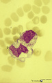 |
Toxic granulation and vacuolation in neutrophilic myelocytes in peripheral blood smear (human) | Stain: May-Grnwald-Giemsa (MGG). The myelocyte (1) and the metamyelocyte (2) show toxic granulation and vacuolation and cytoplasmic swelling. Vacuoles may represent the terminal stage of autophagocytosis. Granules may burst and their contents undergo autophagocytosis in a number of pathologic condit... | Poja Histology Collection - Blood & Bone Marrow Subset | |
| 613 |
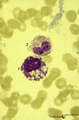 |
Toxic granulation and vacuolization in neutrophilic myelocytes in peripheral blood smear (human) | Stain: May-Grnwald-Giemsa (MGG). The myelocyte (1) and the metamyelocyte (2) show toxic granulation, vacuolisation and cytoplasmic swelling. Toxic granulation is characterized by violet-purple granules of varying size in the cytoplasm; generally admixed with normal pink granules. The phenomenon occu... | Poja Histology Collection - Blood & Bone Marrow Subset | |
| 614 |
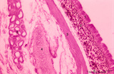 |
Trachea (cat) | Stain: Orcein and eosin. At the top pseudostratified epithelium (1) followed by a thick layer filled with dark-stained elastic fibers (2). Bundle of smooth muscle fibers (3) (note: there is an artifactual space between the elastic layer and the smooth muscle), belonging to the tunica fibro-musculo-... | Tracheal glands; Seromucous glands | Poja Histology Collection - Respiratory System Subset |
| 615 |
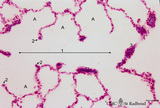 |
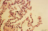
Transition of respiratory bronchiolus into alveolar duct in the lung (human) | Stain: Azan. The alveolar duct (1↔) exhibits an alveolated wall but at a few places (3) bronchiolar characteristics are evident such as cuboidal epithelium covering small areas of smooth muscle and connective tissue. They differ from the characteristic alveolar tips (2↓) of neighboring thin-wall... | Alveolar ducts; Alveolar tips | Poja Histology Collection - Respiratory System Subset |
| 616 |
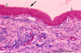 |
Transition surface of vocal cord into laryngeal surface (human, subglottis, higher magnification) | Stain:Stain: Azan. Transition (↓) from stratified squamous epithelium (1) into pseudostratified epithelium (2) in the subglottis region. Blood vessels and the cellularity of the proper lamina is evident. | Olfactory epithelium; Subglottis; Stratified epithelium; Pseudostratified epithelium | Poja Histology Collection - Respiratory System Subset |
| 617 |
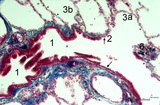 |
Transition terminal bronchiolus - respiratory bronchiolus (human) | Stain: Azan. A longitudinal section shows the transition from the lumen of a terminal bronchiolus (1) into that of a respiratory bronchiolus (2). The epithelium of the terminal bronchiolus is regularly columnar and ciliated. The respiratory bronchiolus has an irregular lining and is covered by low c... | Terminal bronchiolus; Respiratory bronchiolus; Columnar epithelium; Cuboidal epithelium; Alveolar epithelium | Poja Histology Collection - Respiratory System Subset |
| 618 |
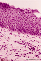 |
Transitional region of epiglottis (human, high magnification) | Stain: Hematoxylin and eosin. Transition of squamous epithelium into pseudostratified epithelium. Note diapedesis of lymphocytes (darker stained rounded cells) through the epithelial barrier (→). The proper lamina presents some lymphocyte infiltration. | Diapedesis | Poja Histology Collection - Respiratory System Subset |
| 619 |
 |
Transitional zone in the middle part of nasal vestibulum (comparable with red zone or vermilion border of lip, human) | Stain: Azan. At the right slightly cornified squamous epithelium (1) with small dermal papillae (2) and numerous blood vessels (3). To the left the transition to pseudostratified epithelium (4). In the submucosa branching seromucous nasal glands with draining ducts (5). | Stratified epithelium; Pseudostratified epithelium; Nasal glands; Seromucous glands | Poja Histology Collection - Respiratory System Subset |
| 620 |
 |
Trophoblast cell in lung alveolar interstitium (human, early midpregnancy) | Stain: Hematoxylin-eosin. It is well known that free circulating trophoblast cells can be found migrated into lung parenchyma during normal pregnancy. In this sample a large trophoblast cell (→) is localised within the normal alveolar interstitium. Alveolar phagocytes (*) are present in the alve... | placenta; trophoblast; lung | Poja Histology Collection - Placenta |
| 621 |
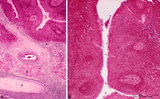 |
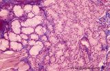
Tubal tonsil (human) | Stain: Azan. The tubal tonsil consists of a collection of lymphoid nodules near the auditory tube opening and forms part of the Waldeyers ring of defense in the nasopharyngeal cavity. This tonsil has fewer crypts (1), and the surface is covered by one to more layered ciliated epithelium (2). The la... | tubal tonsil; nasopharynx | Poja Histology Collection - Lymphatic Tissues and Organs Subset |
| 622 |
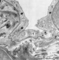 |
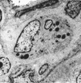
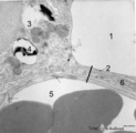
Type I alveolar cell in the lung (human, adult) | Electron microscopy. At the top the alveolar space is lined by type I alveolar cell (1, pneumocyte I). The cytoplasm is well provided with organelles and few electron-dense lysosomes, and pinocytotic vesicles. These pneumocyte I cells cover the interalveolar septa that contain fibroblasts and bundle... | Pneumocyte I; Interstitium | Poja Histology Collection - Respiratory System Subset |
| 623 |
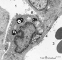 |
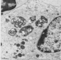
Type II alveolar cell in the lung (rat) | Electron microscopy. At the top the alveolar space (1) is lined by a type II alveolar cell (pneumocyte II) (2) with blunt microvilli. In the cytoplasm the characteristic electron-dense multilamellar bodies (*) and light-stained swollen mitochondria are present. The lamellar bodies are responsible fo... | Pneumocyte I; Pneumocyte II; Alveolar cell type I; Alveolar cell type II; Multilamellar bodies; Interstitial cells | Poja Histology Collection - Respiratory System Subset |
| 624 |
 |
Type II and type I alveolar cells in the lung (human, adult) | Electron microscopy. At the top the alveolar space (1) is lined by a thin cytoplasm (2) of type I alveolar cell (pneumocyte I). At the left side part of a type II alveolar cell (3, pneumocyte II) with electron-dense remnants of two characteristic multilamellar bodies (4) as well as many organelles. ... | Pneumocyte I; Pneumocyte II; Interstitial cell; Alveolar cell type I; Alveolar cell type II | Poja Histology Collection - Respiratory System Subset |
| 625 |
 |
Upper part of nasal septum (dog, higher magnification) | Stain: Hematoxylin and eosin. Stratified squamous epithelium (1) with small papillae (2). The lamina propria contains many small and large thin-walled blood vessels also known as the venous plexus (sinusoids) of Auerbach (3, swell body). Nasal glands (4) with draining ducts (5, larger diameter) and ... | Squamous stratified epithelium; Nasal vestibulum; Venous sinusoids; Venous plexus; Swell bodies | Poja Histology Collection - Respiratory System Subset |