The Health Education Assets Library (HEAL) is a collection of over 22,000 freely available digital materials for health sciences education. The collection is now housed at the University of Utah J. Willard Marriott Digital Library.
TO
Filters: Collection: ehsl_heal
| Title | Description | Subject | Collection | ||
|---|---|---|---|---|---|
| 576 |
 |
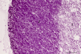
Tertiary villi (human placenta, midpregnancy) | Stain: Hematoxylin - azophloxine. (A) Left a variety of tertiary villi and reddish-stained fibrinoid depositions (1). At (2): the embryonic connective tissue starts to fibrinize. Note syncytiotrophoblast knots (3) or sprouts of several villi .These knots might represent aggregations of degenerati... | placenta; fibrinoid; cytotrophoblast; syncytiotrophoblast | Poja Histology Collection - Placenta |
| 577 |
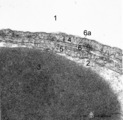 |
The air-blood barrier in the lung (rat) | Electron microscopy (high magnification). The alveolar space (1) and the capillary space (2) (with part of an erythrocyte, 3) are separated from each other respectively by cytoplasm of endothelial cell (5), the common basal lamina (6) with a lamina densa (6a) and the type I alveolar cell (4, pneumo... | Pneumocyte I; Alveolar cell type I | Poja Histology Collection - Respiratory System Subset |
| 578 |
 |
The alveolar tip in lung tissue (human, adult) | Electron microscopy. The lung tip is covered by a flattened alveolar cell type I (↓1) and a similar neighboring bulging one with nucleus (2). In the alveolar tip amorph elastin (5) appears more electron-dense than the collagen fibers (Col) and is interwoven with it. Small foci of calcifications a... | Alveolar tip; Elastoblast | Poja Histology Collection - Respiratory System Subset |
| 579 |
 |
The alveolus and air-blood barrier in the lung (rat) | Electron microscopy. The alveolus (1) is lined by a thin extension (2) of the alveolar epithelial cell type I (2), the pneumocyte I and the thin endothelium (3) of the capillary filled with erythrocytes (4) and blood platelets (5). The thin-walled air-blood barrier (↔) consists of the transition f... | Pneumocyte I; Alveolar cell type I | Poja Histology Collection - Respiratory System Subset |
| 580 |
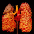 |
The bronchial tree and branches of the pulmonary artery (human, adult, posterior aspect) | Resin corrosion cast of left and right lung. Posterior aspect of lower trachea (1, yellow) with two principal bronchi (yellow) with red-stained pulmonary artery (2) and its branching. The cast of both lung lobes reveals especially the intricate divisions and branching of the bronchial tree (yellow) ... | Macroscopy | Poja Histology Collection - Respiratory System Subset |
| 581 |
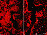 |
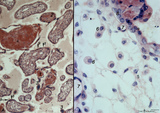
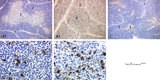
The effect of cyclophosphamide on B cells in spleen (rat) | Stain: Immunofluorescence of Vector red using Mark-1 antibody against B cells. A: Normal rat spleen. (1) the dark, unstained area represents the PALS (periarteriolar lymphatic sheath) filled with T cells. (2) red-stained germinal centre of a B cell follicle that is surrounded by the marginal zone (3... | cyclophosphamide; immunosuppression; immunofluorescence; B lymphocytes | Poja Histology Collection - Lymphatic Tissues and Organs Subset |
| 582 |
 |
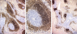
The effect of cyclophosphamide on CD3-thymocytes in thymus (rat) | Stain: Immunoperoxidase staining with diaminobenzidin (DAB) and hematoxylin counterstained on frozen section.. A single injection with cyclophosphamide (CP, 70 mg/ml) induces a transient cortical involution after 4 days, i.e. the dark-blue stained cortex and the lightly stained medulla in normal thy... | cyclophosphamide; CD3 monoclonal antibody; lymphoid tissue ; immunosuppression | Poja Histology Collection - Lymphatic Tissues and Organs Subset |
| 583 |
 |
The effect of cyclophosphamide on splenic B cells (rat) | Stain: Immunoperoxidase staining using diaminobenzidin (DAB)/ hematoxylin counterstained on frozen section of B cells with the antibody Mark-1. The PALS area (1) contains T cells and remains unstained blue. The positively stained B cells (brown) are found in the germinal centres (2) and in the coron... | cyclophosphamide; immunosupression; B lymphocytes; Mark 1 antibody | Poja Histology Collection - Lymphatic Tissues and Organs Subset |
| 584 |
 |
The effect of cyclophosphamide on the CD8-thymocytes in thymus (rat) | Stain: Immunoperoxidase staining with diaminobenzidin (DAB) and hematoxylin counterstained on frozen section. A single injection with cyclophosphamide (CP, 70 mg/ml) induces a transient cortical involution after 4 days, i.e. the darkly stained cortex and the lightly stained medulla in normal thymus ... | cyclophosphamide; CD8 monoclonal antibody; immunosuppression; lymphoid tissue | Poja Histology Collection - Lymphatic Tissues and Organs Subset |
| 585 |
 |
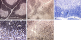
The effect of cyclophosphamide on the resident macrophages in thymus (rat) | Stain: Immunoperoxidase staining with diaminobenzidin (DAB) and hematoxylin counterstained on frozen section. A single injection with cyclophosphamide (CP, 70 mg/ml) induces a transient cortical involution after 4 days. The darkly stained cortex of th normal thymus (A1, A2) decreases its dark stain... | cyclophosphamide; ED1 macrophages ; immunosuppression; lymphoid tissue | Poja Histology Collection - Lymphatic Tissues and Organs Subset |
| 586 |
 |
The effect of cyclophosphamide treatment on the B and T cells in spleen (rat) | Stain: Immunoperoxidase staining using diaminobenzidin (DAB)/ hematoxylin counterstained on frozen section with antibodies to B cells (Mark 1), CD3 and CD8 T cells. (A): B cells in the follicles, germinal centres (2) and corona are stained positive brown, while the PALS area (1) is negative (blue)... | CD3 lymphocytes; CD8 lymphocytes; B lymphocytes; cyclophosphamide | Poja Histology Collection - Lymphatic Tissues and Organs Subset |
| 587 |
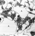 |
The terminal part of the airway: alveoli (dog) | Electron microscopy (low magnification). (A) indicate alveoli; (C) indicate capillaries. (1) type I alveolar cell (pneumocyte I, squamous alveolar cell); (2) type II alveolar cell (pneumocyte II, great alveolar cell). (3) alveolar macrophage. (4) endothelium of capillary. | Pneumocyte I; Pneumocyte II; Alveolar macrophage | Poja Histology Collection - Respiratory System Subset |
| 588 |
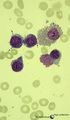 |
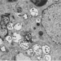
Three basophilic erythroblasts in bone marrow smear (human) | Stain: May-Grnwald-Giemsa (MGG). Three basophilic erythroblasts (1) with intense blue stained cytoplasm, and some so called ears or cytoplasmic projections (arrows). Chromatin strands are thicker than in the proerythroblast. Generally no nucleoli are seen. (2) Damaged or smudged eosinophilic myelocy... | Poja Histology Collection - Blood & Bone Marrow Subset | |
| 589 |
 |
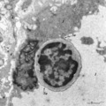
Thymic nurse cell (TNC) (mouse) | Electron microscopy. The thymic nurse cell (TNC) consists of an epithelial reticular cell (type II) enclosing thymocytes. The TNC exists as a sealed structure in situ, i.e. the lymphocytes within the TNC are isolated from the general thymic environment. TNC are located in the cortex, where mature ... | epithelioreticular cell type II; thymic nurse cell | Poja Histology Collection - Lymphatic Tissues and Organs Subset |
| 590 |
 |
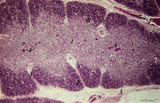
Thymus (human, fetus) | Stain: Silver stain (Gomori). Reticular fibers are demonstrated in the interlobular septa in the thymic cortical area (2) of this lobule. In the medulla (1) the more loosened reticular framework is more distinct. The reticular fibers are produced by the epithelioreticular cells and are particular ... | thymic corpuscle (Hassalls); epithelioreticular cell (ERC); reticular fibers; lymphoid tissue | Poja Histology Collection - Lymphatic Tissues and Organs Subset |
| 591 |
 |
Thymus (human, newborn) | Stain: Hematoxylin & eosin. The thymus is a bilobed lymphoepithelial organ derived as an outgrowth from the third branchial (pharyngeal) pouch, and situated in the anterior mediastinum. Each lobe is divided into multiple lobules by fibrous septa or trabeculae (3). Each lobule consists of an outer co... | epithelioreticular cells (ERC); thymus hormones; Hassalls corpuscle ; lymphoid tissue | Poja Histology Collection - Lymphatic Tissues and Organs Subset |
| 592 |
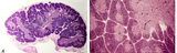 |
Thymus (human, newborn, low and higher magnification) | Stain: Hematoxylin & eosin. The infantile thymus is surrounded by connective tissue capsule (3) from where vascularized interlobular septa (or trabeculae, 3) penetrate into the lobulated organ. Each lobule consists of a darker stained cortex (2) and a lighter stained medulla (1). The medulla has a l... | Zhen; thymus cortex; thymus medulla; Hassall's corpuscle ; Lymphoid tissue | Poja Histology Collection - Lymphatic Tissues and Organs Subset |
| 593 |
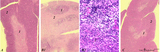 |
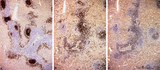
Thymus after cyclophosphamide treatment (rat) | Stain: Hematoxylin & eosin. A single injection with cyclophosphamide (CP, 4 70 mg/ml) induces a transient cortical involution, i.e. inhibition of the cell proliferation and maturation. A: Normal thymus with medulla (1) and cortex (2). B1: Inversion of thymic cortex and medulla 4 to 8 days after CP ... | cyclophosphamide; immunosuppression; involution; lymphoid tissue | Poja Histology Collection - Lymphatic Tissues and Organs Subset |
| 594 |
 |
Thymus cortex (mouse, young adult) | Stain: Hematoxylin. Autoradiography after pulse labeling with tritiated thymidin. Most of the (black) radioactive labeling is found in the outer thymic cortex (3) where pre-T cells divide and subsequently migrate to the lighter stained medulla (1) that consists of a more loosened framework of epith... | thymus cortex; thymus medulla; thymidin labeling; lymphoid tissue | Poja Histology Collection - Lymphatic Tissues and Organs Subset |
| 595 |
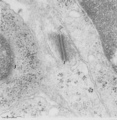 |
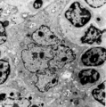
Thymus cortex (mouse, young adult) | Electron microscopy. (1) A well developed desmosome (1) of an epithelioreticular cells type II (TEC2). Apart from few free ribosomes, glycogen granules (2) are present in the electron-light cytoplasm. (3) part of an electron-grey thymocyte with many ribosomes. | epithelioreticular cell type II ; desmosome; lymphoid tissue | Poja Histology Collection - Lymphatic Tissues and Organs Subset |
| 596 |
 |
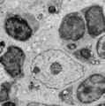
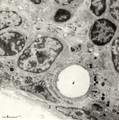
Thymus cortex (mouse, young adult) | Electron microscopy. Surrounded by thymocytes (2) a cortical macrophage (starry-sky macrophage) is seen and shows an electron-light nucleus (N) and a distinct nucleolus. The cell has engulfed two apoptotic thymocytes (1). The cytoplasm also contains small electron-dense lysosomes and myelin figures ... | cortical macrophage; epithelioreticular cell type II ; apoptotic thymocyte; lymphoid tissue | Poja Histology Collection - Lymphatic Tissues and Organs Subset |
| 597 |
 |
Thymus cortex (rat, young adult) | Electron microscopy. Within the thymic cortex a type II epithelioreticular cell or so-called (sometimes multinucleated) thymic nurse cell (TNC) shows a characteristic electron-light nucleus and nucleolus (1). The branches are squeezed between the thymocytes (2). In the cytoplasm a variety of empty a... | MHC class I and II expression; epithelioreticular cell type I and II; thymic nurse cell TNC; lymphoid tissue | Poja Histology Collection - Lymphatic Tissues and Organs Subset |
| 598 |
 |
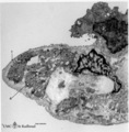
Thymus cortex (rat, young adult) | Electron microscopy. Type I epithelioreticular cells (4) separate connective tissue compartment (capsule, trabeculae, blood vessels) from the thymic parenchyma. At the left the capsule is bordered by a basal lamina (4a) of two projections (4) of type I epithelioreticular cells. Close to them, part o... | lymphoid tissue ; epithelioreticular cell type I; diapedesis | Poja Histology Collection - Lymphatic Tissues and Organs Subset |
| 599 |
 |
Thymus cortex (rat, young adult) | Electron microscopy. Two epithelioreticular cells type II or TEC2 (1) show the characteristic vacuoles (*) partly filled with granules (thymulin, lymphokines). At (--><--) small desmosomes. Apart from the mitochondria electron-dense lysosomal structures are present as well as tonofilaments (1, kera... | epithelioreticular cell II ; desmosome; MHC-II expression; lymphoid tissue | Poja Histology Collection - Lymphatic Tissues and Organs Subset |
| 600 |
 |
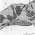
Thymus cortex (rat, young adult) | Electron microscopy. Type I epithelioreticular cells separate connective tissue compartment from the thymic parenchyma. With occludens junctions and desmosomes as barriers they form wide-mesh networks creating specific microenvironments for developing T cells. The extensions of type I cells (5) are... | thymus cortex; epithelioreticular cell type I; epithelioreticular cell type II; lymphoid tissue | Poja Histology Collection - Lymphatic Tissues and Organs Subset |