The Health Education Assets Library (HEAL) is a collection of over 22,000 freely available digital materials for health sciences education. The collection is now housed at the University of Utah J. Willard Marriott Digital Library.
TO
Filters: Collection: ehsl_heal
| Title | Description | Subject | Collection | ||
|---|---|---|---|---|---|
| 526 |
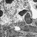 |
Submandibular gland (rat) | Electronmicroscopy. Darkly stained cells around the lumen of an intercalated duct and close to the lightly stained (partly degranulated) serous cells. | oral cavity; seromucous gland; intercalated duct | Poja Histology Collection - Oral Cavity Subset |
| 527 |
 |
Submucosa of trachea (human, adult) | Stain: Goldner trichrome. Mixed tracheal glands (1) of the submucosa (connective tissue light green) and bundles of smooth muscle fibers (2) close to the fibroelastic membrane between the cartilage edges. | Seromucous gland; Tracheal glands | Poja Histology Collection - Respiratory System Subset |
| 528 |
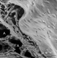 |
Surface of lung pleura (gerbil) | Scanning electron microscopy of visceral pleura. Alveoli (A) and capillaries (c) with scattered erythrocytes (artifactual due to preparation procedures). At (*) connective tissue and in between erythrocytes. The visceral surface is covered by a continuous sheet of flat squamous mesothelial cells (1,... | Visceral pleura; Mesothelium | Poja Histology Collection - Respiratory System Subset |
| 529 |
 |
Surface of olfactory epithelium (rat) | Electron microscopy. Bottom left shows an olfactory bulb (1, vesicle) with cross-sectioned basal bodies. Right side part of the apex of a supporting cell (2) with microvilli (3). Parallel to the surface of the epithelium one long olfactory cilium (4) and several other cross-sectioned ones are detect... | Olfactory epithelium; Olfactory vesicle | Poja Histology Collection - Respiratory System Subset |
| 530 |
 |
Surface of olfactory epithelium in the nose (rat) | Scanning electron microscopy. A carpet of fine long threads of olfactory cilia (*). On top of the carpet broken remnants of cilia clotted due to secretion products. | Olfactory epithelium | Poja Histology Collection - Respiratory System Subset |
| 531 |
 |
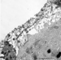
Surface of the nasal septum (rat) | Scanning electron microscopy of anterior part of the nasal septum. The superficial squamous cells of the stratified epithelium are large cells provided with small stubby microvilli. Cell borders are well indicated (↓). | Nasal vestibulum; Stratified epithelium | Poja Histology Collection - Respiratory System Subset |
| 532 |
 |
Surface of trachea epithelium (rat) | Scanning electronmicroscopy. There is an abundancy of cilia with interspersed protrusions (↓) of goblet cells due to apical secretion. | Pseudostratified epithelium | Poja Histology Collection - Respiratory System Subset |
| 533 |
 |
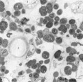
Survey and detail of a peripheral blood smear contaminated with endothelial cells (human) | Stain: May-Grnwald-Giemsa (MGG). Survey (A) and details (B) show aggregates of elongated cells with a large nucleus, mostly situated at the end of a smear between the erythrocytes and granulocytes. These endothelial cells are contaminants derived from blood vessels during puncture. | Poja Histology Collection - Blood & Bone Marrow Subset | |
| 534 |
 |
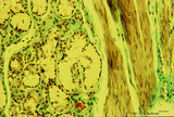
Survey and detail of bone marrow section (human) | Stain: modified hematoxylin and eosin (A, B). The bone marrow consists of bone trabecles or crests (1) covered with osteoclasts and osteoblasts. Between the trabecles bone marrow parenchym (2) consisting of progenitor cells, reticular cells and fat cells (3). Numerous capillaries (B, 4) and enlarged... | Poja Histology Collection - Blood & Bone Marrow Subset | |
| 535 |
 |
Survey and detail of the chorionic plate and intervillous space (human placenta, full-term) | Stain: (A) Perjodic acid-Schiff reaction (PAS); (B) Hematoxylin-azophloxine. (A) At the top the chorionic plate (1) with cross-sections of umbilical vessels (2). At (3) the folded amnion covering the chorionic plate. Ramifications of thicker stem villi (6) demonstrate free-floating terminal villi ... | placenta; chorionic plate; amnion; terminal villi | Poja Histology Collection - Placenta |
| 536 |
 |
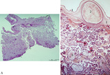
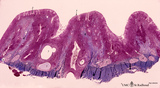
Survey and details of palatine tonsil ('lymphoepithelial tissues', 'gut-associated lymphatic tissue' or GALT) (human) | Stain: Azan. The survey in (A) shows that the palatine tonsil (localized in the lateral wall of the oropharynx) consists of crypts (1) and folds of the surface epithelium (stratified squamous) surrounded by accumulations of lymphoid cells organized in follicles (2). (B): the lining epithelium (5) i... | stratified squamous epithelium | Poja Histology Collection - Lymphatic Tissues and Organs Subset |
| 537 |
 |
Survey frontal section of larynx (human) | Stain: Azan. A pseudostratified epithelium covers the mucosa of the laryngeal cavity and vestibulum. At the vocal fold edge (3) the epithelium appears to be nonkeratinizing squamous. The connective tissue is rigidly attached to the this edge and merges into the vocal ligament (dense elastic) and voc... | Vocal muscle; Vocal ligament; Laryngeal glands; Ventricular fold | Poja Histology Collection - Respiratory System Subset |
| 538 |
 |
Survey nasal septum (dog) | Stain: Hematoxylin and eosin. The nasal septum consists of a cartilage skeleton present as one long (1, cartilago vomeronasalis) and two short plates (2, cartilago septi nasi) with centrally dark-stained areas of chondrocytes. Below, the distal tip is covered with keratinized stratified squamous epi... | Squamous stratified epithelium; Nasal vestibulum; Venous sinusoids; Venous plexus; Swell bodies | Poja Histology Collection - Respiratory System Subset |
| 539 |
 |
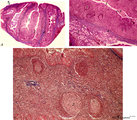
Survey of pharyngeal tonsil ('lymphoepithelial tissues', 'gut-associated lymphatic tissue' or GALT) (human) | Stain Azan. The solitary pharyngeal tonsil is localized in the pharyngeal fornix and belongs to the so-called Waldeyer's ring of pharyngeal lymphatic tissue. A survey shows the folded columnar epithelium (1) with faintly light-stained goblet cells with clefts (2) in between. The lamina propria co... | pharyngeal tonsil; GALT | Poja Histology Collection - Lymphatic Tissues and Organs Subset |
| 540 |
 |
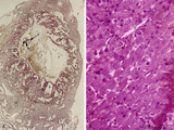
Survey of a centrilobular lung emphysema: section of an lobule (human, adult) | Stain: Hematoxylin and eosin. The enlargement of large areas of the air spaces (X) is evident due to destruction of the walls of alveoli throughout the lobule. | Poja Histology Collection - Respiratory System Subset | |
| 541 |
 |
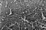
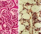
Survey of bone marrow (bone marrow, rabbit) | Electron microscopy. Within the loose reticular tissue a great variety of different stages of young erythrocytes (3, 4) as well as of young granulocytes (5) is present. (2) indicates reticular cells and (2b) phagocytizing reticular cells. (1) fat cells (adipocytes); (6) myeloid cell; (7) capillary. | Poja Histology Collection - Blood & Bone Marrow Subset | |
| 542 |
 |
Survey of bone marrow section (human) | Stain: modified hematoxylin and eosin. In this survey the white spots (1) are fat cells. The marrow is well filled with cells of erythropoietic and myelopoietic series. The very large cells are usually megakaryocytes. Cells with very dark condensed nuclei belong mostly to the erythropoietic series. | Poja Histology Collection - Blood & Bone Marrow Subset | |
| 543 |
 |
Survey of bronchus and lung parenchym (human, adult) | Stain: Azan. (1) Lumen of small bronchus beside a lumen of pulmonary artery (2). Thin plates of hyaline cartilage (3) and connective tissue surrounding (1) and (2). Arrows (→) indicate lung alveoli with black-stained patches of carbon deposits. (4) Lung parenchym with alveoli. | Lung parenchym | Poja Histology Collection - Respiratory System Subset |
| 544 |
 |
Survey of bronchus and lung parenchym (human, adult) | Stain: Azan. Lumina of bifurcated bronchi (1, 2). Thin plates of hyaline cartilage (3) and connective tissue surround the bronchi. Arrows (→) indicate lung alveoli with black-stained patches of carbon deposits. (4) Lung parenchyma with alveoli. | Lung parenchym | Poja Histology Collection - Respiratory System Subset |
| 545 |
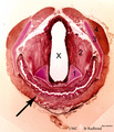 |
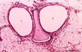
Survey of cross-section through larynx-esophagus (human) | Stain: Hematoxylin and bordeaux light. X = lumen of larynx (middle part); ↑ = esophagus (below) with an undulating mucosa covered with squamous epithelium. Elastic arythenoid cartilage (1); thyreoarythenoid muscle (2); thyroid cartilage (3); hyoid muscle (4). | Arythenoid cartilages | Poja Histology Collection - Respiratory System Subset |
| 546 |
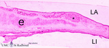 |
Survey of epiglottis (human) | Stain: Azan. Centrally light-stained elastic cartilage (e). Lingual side (LI) is covered with squamous epithelium. At the laryngeal side (LA) the squamous epithelium usually turns into respiratory epithelium with seromucous laryngeal glands (*). | Laryngeal gland; Oral cavity | Poja Histology Collection - Respiratory System Subset |
| 547 |
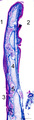 |
Survey of epiglottis (human) | Stain: Azan. Centrally light-stained elastic cartilage (4). Lingual side (2) is covered with squamous epithelium. At the laryngeal side (1) the squamous epithelium usually turns into respiratory epithelium with seromucous laryngeal glands (5). Note also lymphoid aggregations in this area (3 →). | Oral cavity; Laryngeal glands | Poja Histology Collection - Respiratory System Subset |
| 548 |
 |
Survey of implanted blastocyst (human, ca. 3 weeks-old embryo) and detail of advanced stage of decidua cells (human, mid pregnancy) | Stain: Hematoxylin-eosin (A, left) and Azan (B, right). (A) Left: section through a slightly damaged implantation site shows at (1) the uterine lumen and (2) the decidua capsularis while at (3) several uterine glands are visible. At arrow (4) the amniotic cavity, (5) points to embryonic shield and... | placenta; uterus; syncytiotrophoblast; implantation; blastocyst | Poja Histology Collection - Placenta |
| 549 |
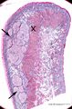 |
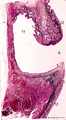
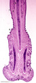
Survey of lip (human) | Stain: Azan. Arrows: labial glands in submucosa (mucous inner surface with non-keratinized epithelium). X: orbicularis oris muscle. Right side = skin surface (keratinized epithelium). Top = transitional zone (red zone) with slightly keratinized epithelium. | oral cavity; red zone; labial glands | Poja Histology Collection - Oral Cavity Subset |
| 550 |
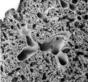 |
Survey of lung parenchym (gerbil) | Scanning electron microscopy. The photograph shows (1) bifurcation of several bronchi in the lung parenchym with alveoli (2). The long arrows indicate the route of alveolar ducts (4 ↕ length, and cross 4↑). Pulmonary vessels (3). | Lung parenchym; Pulmonary vessels | Poja Histology Collection - Respiratory System Subset |