The Health Education Assets Library (HEAL) is a collection of over 22,000 freely available digital materials for health sciences education. The collection is now housed at the University of Utah J. Willard Marriott Digital Library.
TO
Filters: Collection: ehsl_heal
| Title | Description | Subject | Collection | ||
|---|---|---|---|---|---|
| 476 |
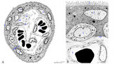 |
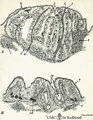
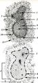
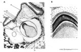
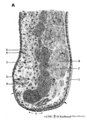
Scheme of electron microscopy of tertiary villus (human placenta, midpregnancy) | (See also POJA-L1227). (A) Survey and (B) detail of a tertiary placental villus lined by the multinucleated syncytiotrophoblast cell (1) (STC). The apex shows protrusions and extensive microvilli of varying sizes (brushborder, 2). Nuclei are sectioned at different levels and the cytoplasm contains a... | placenta; tertiary villi; placental barrier; electron microscopy | Poja Histology Collection - Placenta |
| 477 |
 |
Scheme of filiform and fungiform papillae of the tongue - human, adult | 5. filiform papillae; 6. fungiform papillae; 8. locally cornification on top of filiform papillae; 9. lingual muscle; 10. lingual glands | oral cavity; papillae | Poja Histology Collection - Oral Cavity Subset |
| 478 |
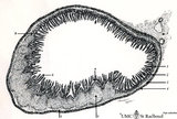 |
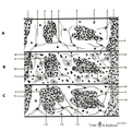
Scheme of ileum with Peyers patches (gut-associated lymphatic tissue or GALT) (dog) | Survey ileum (see also Digestive System: Ileum) A large amount of non-encapsulated diffuse lymphatic tissue or mucosa-associated lymphatic tissue (MALT) is located in the subepithelial lamina propria (e.g. respiratory passages, genitourinary tract). The gut-associated lymphatic tissues (GALT) are ... | GALT; follicle; scheme; germinal center | Poja Histology Collection - Lymphatic Tissues and Organs Subset |
| 479 |
 |
Scheme of lingual tonsil ('lymphoepithelial tissue') | Lingual tonsil (consisting of the accumulation of folliculi linguales). The root of the tongue contains invaginations or crypts or narrow caverns (3). In these crypts the ducts of the mucous glands (8) end up. The crypts are lined by multilayered, non-keratinizing squamous epithelium (2) and are sur... | lingual tonsil; scheme; germinal center; follicle | Poja Histology Collection - Lymphatic Tissues and Organs Subset |
| 480 |
 |
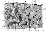
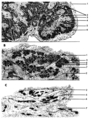
Scheme of lymph node (human) | Lymph node: A. survey; B. detail subcapsular (or marginal) sinus 1. adipose tissue; 2. capsule; 3. afferent lymph vessel with valves (B); 4. subcapsular (or marginal) sinus; 5. germinal centre of a secondary lymphatic nodule (or follicle); 6. crescent or mantle zone indicated by cap of per... | cortex; scheme; paracortex; germinal center | Poja Histology Collection - Lymphatic Tissues and Organs Subset |
| 481 |
 |
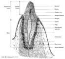
Scheme of palatine and pharyngeal tonsils ('lymphoepithelial tissues') (human) | The palatine tonsils are covered by the multilayered, non-keratinizing squamous epithelium of the oral cavity (A-1). In contrast to the palatine and lingual tonsils, the epipharyngeal tonsil has a multilayered ciliated epithelium (respiratory-like epithelium, B-9). The Waldeyer's tonsillar ring i... | germinal center; scheme; follicle | Poja Histology Collection - Lymphatic Tissues and Organs Subset |
| 482 |
 |
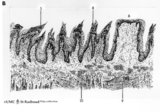
Scheme of peripheral lung parenchym (human, adult) | (1) respiratory bronchiolus; (2) alveolar duct; (3) alveolar sac is a virtual sac formed by several alveoli, but continuous with the alveolar duct; (4) alveoli which end in small tips (↓) with elastin interwoven with collagen. | Alveolar duct | Poja Histology Collection - Respiratory System Subset |
| 483 |
 |
Scheme of placental tissues (human) | See the http://www.healcentral.org/http://library.med.utah.edu/healassets/content/collections/poja_placenta/POJA-L1227-FemReprOrg-Placenta-scheme+introduction Placenta.pdf>detailed Legends for this slide (PDF file). | placenta; uterus; chorionic villi; decidua; trophoblast; cytotrophoblast | Poja Histology Collection - Placenta |
| 484 |
 |
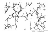
Scheme of reticular tissue in lymph node | Lymph node stroma does not contain stromal cells but instead the reticular meshwork is build by reticular cell types, macrophages and lymphoid cells and myeloid cells. Route (A) in the scheme represents morphological differentiation path from B lymphocytes to plasma cells. 1 endothelial ce... | reticular tissue; scheme; follicle; germinal center | Poja Histology Collection - Lymphatic Tissues and Organs Subset |
| 485 |
 |
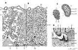
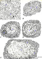
Scheme of secondary lymphatic nodule in spleen (human) | A. histological impression of a follicle or nodule B. outline of impression 1. pulpa artery; 2. central artery ; 3. lymphatic sheath; 4. lymphatic nodule; 5. thymus-dependent area (periarteriolar lymphatic sheath or PALS), composed of Th subset lymphocytes; 6. mantle zone with mainly nave B l... | white pulp; marginal zone; antigen presenting cell (APC) | Poja Histology Collection - Lymphatic Tissues and Organs Subset |
| 486 |
 |
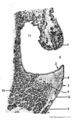
Scheme of the epiglottis (human, adult) | The laryngeal side of the epiglottis (1) is covered with respiratory epithelium, while the lingual side (2) is similar to the oral cavity epithelium (squamous type). The transitional zone to respiratory epithelium is marked by (3). The scaffold consists of elastic cartilage (4). Seromucous laryngea... | Laryngeal glands; Oral cavity | Poja Histology Collection - Respiratory System Subset |
| 487 |
 |
Scheme of the medullar changes in lymph node after antigen stimulation | Upon antigenic stimulation the reticular cells that line the medullar sinusoids start to phagocytise the antigens (i.e. infective agents) and change from small tiny stellate cells into swollen large cells due to phagocytosis. They detach from the medullar sinus wall. After ten days the situation has... | follicle; antigen stimulation; scheme; germinal center | Poja Histology Collection - Lymphatic Tissues and Organs Subset |
| 488 |
 |
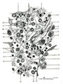
Scheme of the spleen (human) | Spleen: A. general diagram; B. adult; C. senium 1. capsule of dense irregular connective tissue with few elastic and smooth muscle fibers (it varies with the species); 2. trabecula (septum); 3. trabecular artery derived from the splenic artery; 4. trabecular vein; 5. when the pulpa artery ... | white pulp; red pulp | Poja Histology Collection - Lymphatic Tissues and Organs Subset |
| 489 |
 |
Scheme of the thymus during ageing (human) | Physiological changes in the course of human ageing lead to almost complete degeneration of the thymus (age involution). Both cortex and medulla become depleted of lymphocytes, and the distinction between both layers gradually becomes less. The remaining reticular meshwork of connective tissue is gr... | thymus involution; thymus development; thymus age; lymphoid tissue | Poja Histology Collection - Lymphatic Tissues and Organs Subset |
| 490 |
 |
Scheme of the thymus medulla (human) | A-B: medullar epithelioreticular cells; C-D-E: development stages of thymic corpuscles (or Hassall bodies) (1): branching (-->) epithelioreticular cells in the medulla forming a meshwork (cytoreticulum) that normally is populated by thymocytes; (2): specialized epithelioreticular cells (2) often... | thymic corpuscle; Hassall ; epithelioreticular cells ; lymphoid tissue | Poja Histology Collection - Lymphatic Tissues and Organs Subset |
| 491 |
 |
Scheme of tongue papillae and foliate papillae | A. Circumvallate papillae (tongue, human, adult) magnification x 35 objective. B. Foliate papillae (tongue, rabbit) magnification x 85 objective. C. Taste bud in foliate papilla (tongue, rabbit) magnification x 750 objective. 1. (circum)vallate papilla; 2. non-keratinized stratified squamous epit... | oral cavity; von Ebner; circumvallate papillae; foliate papillae | Poja Histology Collection - Oral Cavity Subset |
| 492 |
 |
Scheme of tongue papillae and taste buds | B. Scheme of foliate papillae of the tongue (rabbit) magnification x 85 objective. C. Scheme of taste bud in foliate papilla of the tongue (rabbit). 2. non-keratinized stratified squamous epithelium; 3. taste bud; 4. serous gland (von Ebner); 5. draining duct (of serous gland); 7. foliate papil... | oral cavity; von Ebner; foliate papillae | Poja Histology Collection - Oral Cavity Subset |
| 493 |
 |
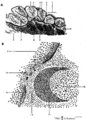
Scheme of tooth development (human) | A. Survey of tooth germ (bell stage, embryo) magnification x 35 objective. B. Formation of enamel and dentin (bell stage, embryo) magnification x 350 objective. 1. pulp organ; 2. odontoblasts; 3. mantle predentin; 4. dentin; 5. enamel; 6. initial enamel; 7. inner dental epithelium (ameloblasts);... | oral cavity; dental lamina; bell stage | Poja Histology Collection - Oral Cavity Subset |
| 494 |
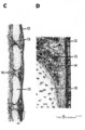 |
Scheme of trachea | C. Trachea (human, adult) magnification x 7.5 objective). D. Trachea (human, adult, detail of rectangle in C) magnification x 85 objective. (12) pseudostratified ciliated epithelium; (13) seromucous tracheal glands; (14) condensed layer of elastic fibers at the border of lamina propria and submu... | Pseudostratified epithelium ; Fibroelastic membrane; Adventitia | Poja Histology Collection - Respiratory System Subset |
| 495 |
 |
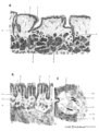
Scheme of trachea (human, adult) | On top pseudostratified ciliated epithelium (1) with goblet cells followed by a condensed layer of elastic fibres (2) and the transition (*) to seromucous tracheal glands (3) close to the edge of the perichondrium (4) of hyaline cartilage (5). | Pseudostratified epithelium ; Perichondrium | Poja Histology Collection - Respiratory System Subset |
| 496 |
 |
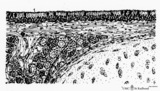
Scheme survey dorsal surface of tongue - human, adult | 1. foliate papillae (rudimentary in human); 2. circumvallate papillae; 3. foramen cecum; 4. palatine tonsils; 5. filiform papillae; 6. fungiform papillae; 7. epiglottis; 11. lingual tonsils | oral cavity | Poja Histology Collection - Oral Cavity Subset |
| 497 |
 |
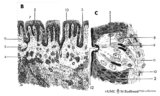
Scheme survey frontal section of larynx (human) | Stain: Azan. A pseudostratified epithelium covers the mucosa of the laryngeal cavity and vestibulum. At the vocal fold edge (3) the epithelium appears to be nonkeratinizing squamous. The connective tissue is rigidly attached to this edge and merges into the vocal ligament (dense elastic) and vocal m... | Vocal ligament; Laryngeal glands; Ventricular fold | Poja Histology Collection - Respiratory System Subset |
| 498 |
 |
Scheme survey lip - human, adult | 1. red (transitional) zone; 2. keratinized stratified squamous epithelium (outer side); 3. non-keratinized stratified squamous epithelium (inner side); 4. hair shaft; 5. hair follicle; 6. sebaceous gland; 7. cross-section of circumoral muscle; 8. labial glands with draining ducts; ... | oral cavity | Poja Histology Collection - Oral Cavity Subset |
| 499 |
 |
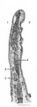
Scheme survey tooth (longitudinal section; human; canine) | Canine tooth in osseous alveolus. | oral cavity; pulp canal | Poja Histology Collection - Oral Cavity Subset |
| 500 |
 |
Scheme survey uvula - soft palate, human, adult | 1. nasal side - pseudostratified columnar ciliated epithelium (respiratory epithelium); 2. oral side - non-keratinized stratified squamous epithelium; 3. mixed glands (pharyngeal glands); 4. uvular muscle; 5. mucous palatine glands | oral cavity | Poja Histology Collection - Oral Cavity Subset |