John A. Moran Eye Center Neuro-Ophthalmology Collection: A variety of lectures, videos and images relating to topics in Neuro-Ophthalmology created by faculty at the Moran Eye Center, University of Utah, in Salt Lake City.
NOVEL: https://novel.utah.edu/
TO
Filters: Collection: ehsl_novel_jmec
| Identifier | Title | Description | Subject | ||
|---|---|---|---|---|---|
| 26 |
 |
1-5 | Third Nerve Palsy, Pupil Involving | Example of patient with third nerve palsy. Left eye shows pupilary involvement. Left eye doesn't immediately duct, but abducts well, with impaired superduction. Secondary and primary deviations are demonstrated. Anisocoria is more prominent when light is on, showing a parasympathetic defect to the p... | Pupil; Third Nerve Palsy; Third Nerve Dysfunction |
| 27 |
 |
1-6 | Aberrant Regeneration of Third Nerve, Bilaterally (1 degree OD, 2 Digrees OS) | Example of patient with bilateral aberrancy of the third nerve. Shows lids popping up (synkinetic) with adduction. Patient had bilateral internal carotid artery aneurisms with third nerve compression. | Bilateral Aberrant Regeneration of Third Nerve; Third Nerve Dysfunction |
| 28 |
 |
1-7 | Aberrant Regeneration of the Third and Sixth Nerves | Aberrant Regeneration; Third and Sixth Nerves; Third Nerve Palsy | |
| 29 |
 |
1-8 | Internuclear Ophthalmoplegia (2 Examples) | Two examples of patients with internuclear ophthalmoplegia. First patient has a right internuclear ophthalmoplegia. Patient had subacute bacterial endocarditis with a bacterial abscess in the brain stem. Ductions and gaze to the right look good, but when gazing to the left, the right eye does not ad... | Internuclear Ophthalmoplegia; Abducting Nystagmus |
| 30 |
 |
1-9 | Ocular Lateropulsion (Wallenberg's Syndrome) | Example of patient with ocular lateropulsion. Patient also has central Horner syndrome and nystagmus in right gaze. When shifting gaze back to forward, eyes overshoot their mark. Eyes laterally deviate to the right upon opening. | Ocular Lateropulsion; Wallenberg's Syndrome; Lateropulsion; Lateral Medullary Syndrome; Posterior Inferior Cerebellar Artery; Wallenberg Syndrome |
| 31 |
 |
2-1 | Congenital Nystagmus | Example of patients with congenital nystagmus. First patient's nystagmus are mostly jerk and not pendular. Second patient's nystagmus are mostly pendular. Both patients show a uniform horizontal oscillation. Second patient also shows differences in frequency of oscillations depending on gaze, includ... | Congenital Nystagmus |
| 32 |
 |
2-10 | Binocular Pendular Nystagmus | Example of a patient with binocular pendular nystagmus. Patient has somewhat dissociated nystagmus, with nystagmus seen more prominently in the left eye. Patient shows an occasional jerk nystagmus to the right in the right eye. Left eye oscillations are mostly pendular. | Binocular Pendular Nystagmus; Pendular Nystagmus; Acquired Pendular Nystagmus |
| 33 |
 |
2-11 | Brun's Nystagmus | Observation of patient with Brun's Nystagmus. Shows patient gazing to the right and the nystagmus beating in the direction of the gaze. | Brun's Nystagmus; Cyclical |
| 34 |
 |
2-12 | Periodic Alternating Nystagmus | Example of a patient with periodic alternating nystagmus, showing an alternation between left-beats and right-beats as the patient maintains forward gaze. Nystagmus maintain horizontal direction regardless of position of gaze. | Periodic Alternating Nystagmus |
| 35 |
|
2-13 | Rotary Nystagmus | Example of a patient with rotary nystagmus, showing occasional counterclockwise rotary movements of both eyes. Seen more in intrinsic disorders of the brainstem. | Rotary (Torsional) Nystagmus |
| 36 |
 |
2-14 | Dissociated Nystagmus | Example of a patient with dissociated nystagmus. Demonstrates difference in movements between each eye. | Dissociated Nystagmus |
| 37 |
 |
2-15 | Vestibular Nystagmus | Discussion of vestibular nystagmus. Seen with peripheral disorders and central disorders, and in two varieties: spontaneous and positional. Horizontal jerk with small amplitude. | Vestibular Nystagmus; Jerk Nystagmus; Peripheral Vestibular Nystagmus; Positional Nystagmus |
| 38 |
 |
2-16 | Physiologic (End-Gaze) Nystagmus | Demonstration of physiological nystagmus, where oscillations do not represent pathology, but occur when the patient's gaze is drawn too far laterally. | End-Gaze Nystagmus; Physiologic Nystagmus |
| 39 |
|
2-17 | Ocular Flutter | Two examples of patients, the first with rotary, flutter-like movements, but not ocular flutter, and the second with genuine ocular flutter. Discussion of difference between ocular flutter and nystagmus, and how to elicit ocular flutter. | Ocular Flutter |
| 40 |
 |
2-19 | Superior Oblique Myokymia | Example of patients with superior oblique myokymia, a saccadic intrusion. First patient is seen to have intermittent, intorting movements with superimposed slight vertical deviations in right eye. Discussion of disorder as benign, but frequently disabling, as patients experience episodes of diplopia... | Superior Oblique Myokymia; Third Nerve Palsy |
| 41 |
|
2-2 | Latent Nystagmus | Example of a patient with latent nystagmus. Demonstrates a lack of oscillations in forward gaze, followed by the occlusion of each eye, showing how this generates a jerking oscillation in the non-occluded eye away from the occluded eye. | Latent Nystagmus; Fusional Maldevelopment Nystagmus Syndrome |
| 42 |
 |
2-20 | Square Wave Jerks | Example of patient with square wave jerks. Discussion of difference between square wave jerks (saccadic oscillations) and horizontal nystagmus. | Square Wave Jerks |
| 43 |
 |
2-21 | Convergence Retraction Nystagmus (Parinaud's Syndrome) | Examples of patients with convergence retraction nystagmus. Shows saccadic oscillations in patients looking upwards and following downwards moving targets. Also shows a side-view of the retracting movements of the globes. | Convergence Retraction Nystagmus; Parinaud's Syndrome; Dorsal Midbrain Syndrome; Lid Retraction |
| 44 |
 |
2-22 | Voluntary Nystagmus | Example of patient with voluntary nystagmus. Discussion of how a lack of uniform, patterned movement of the eyes along with associated lid movements suggests that activity is voluntary. | Voluntary Nystagmus; Voluntary Flutter |
| 45 |
 |
2-3 | Spasmus Nutans | Example of patient with spasmus nutans. Discussion of characteristics of this disorder, such as dissociated or monocular nystagmus, abnormal head position, and to-and-fro head oscillation. Sometimes an eccentric gaze is seen as well (as in patient). Patient has a monocular horizontal nystagmus in th... | Spasmus Nutans |
| 46 |
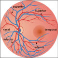 |
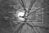
2-37a | 2-37a - Vascular Features | When looking at the disc, the central retinal artery and vein should be visible. The central retinal artery is usually slightly narrower than the vein. When the central retinal artery goes though the lamina cribrosa, the artery becomes smaller because of diminution of the muscular layer and loss of ... | Vascular Features |
| 47 |
 |
2-37b | 2-37b - Vascular Features | When looking at the disc, the central retinal artery and vein should be visible. The central retinal artery is usually slightly narrower than the vein. When the central retinal artery goes though the lamina cribrosa, the artery becomes smaller because of diminution of the muscular layer and loss of ... | Vascular Features |
| 48 |
|
2-4 | See-saw Nystagmus | Example of a patient with see-saw nystagmus, showing how one eye elevates as the other depresses, with the elevating eye intorting as the depressing eye extorts. Shows vertical oscillations with pendular waveforms. Suggests a large structural lesion in the pericellar region (associated with bi-tempo... | See-saw Nystagmus |
| 49 |
 |
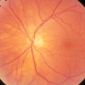
2-4a | 2-4a - Disc Anatomy | The optic disc appearance is determined by: the size of the eye, the size of the scleral canal, how the nerve is inserted into the globe, the appearance of the lamina cribrosa, where myelination stops, and what is left behind in normal development. Even though this is a disc with a very large cup, i... | Disc Anatomy |
| 50 |
 |
2-5 | Upbeat Nystagmus | Example of a patient with upbeat nystagmus. Shows vertical jerk nystagmus with fast phases in the up direction. Localizes to brain stem, and occurs with strokes, demyelination, and tumors. | Upbeat Nystagmus; Blepharospasm |