John A. Moran Eye Center Neuro-Ophthalmology Collection: A variety of lectures, videos and images relating to topics in Neuro-Ophthalmology created by faculty at the Moran Eye Center, University of Utah, in Salt Lake City.
NOVEL: https://novel.utah.edu/
TO
| Title | Description | Type | ||
|---|---|---|---|---|
| 26 |
 |
MELAS and RP | MELAS; Mitochondrial Encephalopathy with Lactic Acidosis, Stroke and Pigmentary Changes in retina-associated with a retinal dystrophy. This 53 year old man had seizures, encephalopathy and lactic acidosis typical of MELAS. His fundus examination showed granularity and some slight pigmentary changes ... | Text |
| 27 |
 |
Silent Sinus Syndrome | Silent sinus syndrome (SSS) is characterized by spontaneous and progressive unilateral enophthalmos. | |
| 28 |
 |
Benign Episodic Unilateral Mydriasis | Presentation covering benign episodic mydriasis. | Text |
| 29 |
 |
Near Reflex and Accomodation | Description of testing the near reflex and accomodation. | |
| 30 |
 |
Optic Disc: Anatomy, Variants, Unusual discs | Discussion of viewing the optic disc. Includes development of direct ophthalmoscope. Covers normal optic disc and nerve fiber; nerve fiber loss and defects; cilioretinal arteries; venous anomolies; papilledema; pseudopapilledema; myopic disc; hyperoptic disc; little red discs; megallopapilla; myelin... | Text |
| 31 |
 |
Normal Optic Disc | Overview of the structure and function of the normal optic disc. | Text |
| 32 |
 |
Stages of Papilledema | Text | |
| 33 |
 |
Documenting the Neuro-ophthalmic Patient: External Photography | Description of documenting the neuro-ophthalmic patient using external photography. This covers pupils and extra ocular muscles. | |
| 34 |
 |
The 3 Step Test: Looking for a 4th Nerve Palsy | Description of the three step test (3 step test) used when looking for a 4th nerve palsy. | Text |
| 35 |
 |
Tunnel Vision on Tangent Screen Testing | Description of tunnel vision and tangent screen testing. | |
| 36 |
 |
Spiral and Stellate Visual Fields Non-physiologic Variants | Description of testing the spiral and stellate visual fields. | |
| 37 |
 |
Clover-leaf Visual Field Defects | Description of clover-leaf visual field defects. | |
| 38 |
 |
Tangent Screen Testing Visual Field | Description of tangent screen testing. | |
| 39 |
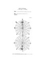 |
Tangent Screen Recording Chart | The tangent screen recording chart. | |
| 40 |
 |
The Electroretinogram and Electro-oculogram: Clinical Applications | The global or full-field electroretinogram (ERG) is a mass electrical response of the retina to photic stimulation. The ERG is a test used worldwide to assess the status of the retina in eye diseases in human patients and in laboratory animals used as models of retinal disease. | Text |
| 41 |
 |
Visually Evoked Potentials | Detailed explanation of visually evoked potentials. The terms visually evoked potential (VEP), visually evoked response (VER) and visually evoked cortical potential (VECP) are equivalent. They refer to electrical potentials, initiated by brief visual stimuli, which are recorded from the scalp overl... | Text |
| 42 |
 |
The Electro-oculogram: Clinical Applications | The electrooculogram measures the potential that exists between the cornea and Bruch's membrane at the back of the eye. The potential produces a dipole field with the cornea approximately 5 millivolts positive compared to the back of the eye, in a normally illuminated room. Although the origin of th... | Text |
| 43 |
 |
Fluoresein Angiography | Comprehensive description of using fluoresein angiography in examinations. |