The Walsh Society Annual Meeting Archives: Proceedings of the annual meeting of the Walsh Society, which is now part of the North American Neuro-Ophthalmology Association (NANOS) Annual Meeting. Contains records from the first meeting in 1969, through the present.
NOVEL: https://novel.utah.edu/
TO
| Title | Creator | History | ||
|---|---|---|---|---|
| 26 |
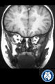 |
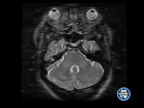
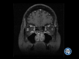
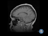
Highly Impossible (Follow-Up MRI Coronal) | William A. Fletcher, MD, Departments of Clinical Neurosciences & Surgery, University of Calgary | A 4-year old male with 1 1/2-year history of visual loss OU. Previous history significant for megacephaly. |
| 27 |
 |
Painful Red Eye in a 69-Year Old Female With Sensorineural Hearing Loss (Presentation Video) | Engelbert, Micahel | A 63-year old female with a 6-year history of ulcerative colitis and erythema nodosum. Previous history significant for bilateral hearing loss, diffuse headache preceded by intermittent vertigo and tinnitus. |
| 28 |
 |
Do You See Mike? (Presentation Video) | Deborah I. Friedman, MD, MPH, Professor, Neurology & Neurotherapeutics, University of Texas Southwestern | A 59-year old male with progressive right facial numbness, right facial pain, diplopia, headache and a 10-pound weight loss. |
| 29 |
 |
A 28 Year-Old Man with Visual Loss and Pain on Eye Movement (Presentation video) | Mark L. Moster, MD, Thomas Jefferson University | A 28-year old male with loss of vision OS associated with soreness on eye movement over an 8-day period. |
| 30 |
 |
A Lebanese Woman with Progressive Visual Loss; presentation video | Gregory P. Van Stavern, MD, Associate Professor, Ophthalmology & Visual Sciences and Neurology, Washington University School of Medicine | A 69-year old female with progressive loss of vision OD. |
| 31 |
 |
Star Light, Star Bright (Presentation Video) | Shannon C. Lynch, MD, University of Nebraska Medical Center | An 83-year female with ptosis and binocular oblique diplopia. |
| 32 |
 |
Oh Great, I Get to Follow the Post-Op Homonymous Hemianopsia (Presentation Video) | Robert F. Saul, MD, Virginia Tech Carilion | A 32-year old female with a right homonymous hemianopsia and 1-month history of severe headache. |
| 33 |
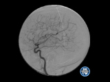 |
When All Else Fails, Lay on Hands (Angiograms RICA) | Roger E. Turbin, MD, Rutgers New Jersey Medical School | A 57-year old female with an 8-month history of photopsia and dark spots OS 1 month after second renal transplant. Renal failure attributed to biopsy-proven renal sarcoidosis. |
| 34 |
 |
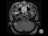
When All Else Fails, Lay on Hands (MRI Axial) | Roger E. Turbin, MD, Rutgers New Jersey Medical School | A 57-year old female with an 8-month history of photopsia and dark spots OS 1 month after second renal transplant. Renal failure attributed to biopsy-proven renal sarcoidosis. |
| 35 |
 |
When All Else Fails, Lay on Hands (MR Contaxial) | Roger E. Turbin, MD, Rutgers New Jersey Medical School | A 57-year old female with an 8-month history of photopsia and dark spots OS 1 month after second renal transplant. Renal failure attributed to biopsy-proven renal sarcoidosis. |
| 36 |
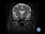 |
When All Else Fails, Lay on Hands (MR Contaxial) | Roger E. Turbin, MD, Rutgers New Jersey Medical School | A 57-year old female with an 8-month history of photopsia and dark spots OS 1 month after second renal transplant. Renal failure attributed to biopsy-proven renal sarcoidosis. |
| 37 |
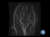 |
When All Else Fails, Lay on Hands (MRI MRV) | Roger E. Turbin, MD, Rutgers New Jersey Medical School | A 57-year old female with an 8-month history of photopsia and dark spots OS 1 month after second renal transplant. Renal failure attributed to biopsy-proven renal sarcoidosis. |
| 38 |
 |
When All Else Fails, Lay on Hands (MRI Contcoron) | Roger E. Turbin, MD, Rutgers New Jersey Medical School | A 57-year old female with an 8-month history of photopsia and dark spots OS 1 month after second renal transplant. Renal failure attributed to biopsy-proven renal sarcoidosis. |
| 39 |
 |
When All Else Fails, Lay on Hands (MRI Sagittal) | Roger E. Turbin, MD, Rutgers New Jersey Medical School | A 57-year old female with an 8-month history of photopsia and dark spots OS 1 month after second renal transplant. Renal failure attributed to biopsy-proven renal sarcoidosis. |
| 40 |
 |
When All Else Fails, Lay on Hands (Presentation Video) | Roger E. Turbin, MD, Rutgers New Jersey Medical School | A 57-year old female with an 8-month history of photopsia and dark spots OS 1 month after second renal transplant. Renal failure attributed to biopsy-proven renal sarcoidosis. |
| 41 |
 |
Diagnosis of Inclusion (Presentation Video) | Iris Ben-Bassat Mizrachi, MD, The Goldschleger Eye Institute | A 50-year old female with a 2-day history of headache and fever found unresponsive at home. |
| 42 |
 |
An Unusual Origin of Balint's Syndrome (Presentation Video) | Iris Ben-Bassat Mizrachi, MD, The Goldschleger Eye Institute | A 50-year old female with progressive visual problems and cognitive deterioration over a 3-month period. |
| 43 |
 |
Horrible Hallucinations (Presentation Video) | Melissa W. Ko, MD, FAAN, Upstate University Hospital | A 34-year old female with a history of irritable bowel and polycystic ovarian syndrome. |
| 44 |
 |
A Mercurial Course (Presentation Video) | Melissa W. Ko, MD, FAAN, Upstate University Hospital | A 61-year old male with a 9-month history of progressive diplopia, dysarthria, ataxic gait, worsening memory impairment, anxiety depression, sleep disturbances and a 30-pound loss of weight. |
| 45 |
 |
Confusing Diplopia (Presentation Video) | Dan Boghen, MD, FRCP(C), Neuro-Ophthalmology Section, Neurology Service, Hôtel-Dieu Hospital, Quebec | A 45-year old female with diplopia. |
| 46 |
 |
Oh My GAD!! Something Else? | Olwen Murphy, Kemar Green, John Probasco, Daniel Gold | A 43 -year-old man presented with oscillopsia, dizziness, binocular vertical diplopia, and gait difficulties. He reported a six-month history of abdominal pain, anorexia and 50-pound weight loss, and a 3-month history of mood and cognitive changes. Torsional nystagmus, a left 4th nerve palsy (NP), u... |
| 47 |
 |
Never Too Young or Too Old | Bart Chwalisz, Laurel Tainsh, Mary Maher, Samantha Champion, Shuhei Nishiyima, Michael Levy | An 81-year-old woman with history of ocular myasthenia gravis presented with sequential bilateral vision loss. Six days before presentation, she discovered that vision of her left eye was reduced to light perception. She did not have any eye pain, pain with eye movement, headache, scalp tenderness, ... |
| 48 |
 |
Gone but Not Forgotten | Jonathan Micieli, Adriana Krizova, Walter Montanera | A 52 -year-old healthy woman presented with a 1-week history of blurred vision and "soreness" in her left eye. Neuro-ophthalmic examination revealed a visual acuity of 20/20 OD, 20/40 OS, left RAPD and left superior arcuate defect on Humphrey visual field testing. Dilated fundus examination demonstr... |
| 49 |
 |
A Small Leak Will Sink a Great Ship | Konstantinos Douglas, Vivian Paraskevi Douglas, Cameron Sadegh, David Chow, Sigurros Davidsdottir, Ganesh Shankar, Bradley Buchbinder, Bart Chwalisz | A 68 -year-old right -handed man presented for evaluation of progressive and fluctuating polyopia, gait instability and cognitive changes of about 14 months. Prior medical history was notable for Crohn's disease in remission, atrial fibrillation, a single episode of aseptic meningitis 12 years prior... |
| 50 |
 |
Orbiting a Diagnosis | Daniel Liebman, Daniel Lefebvre, Emily Tam, Marie Lithgow, Bart Chwalisz, Eric Gaier, Joseph Kane | A 75 -year-old male with a history of chronic/small lymphocytic leukemia (CLL/SLL) presented for one day of left retro-orbital headache, painful eye movements, eyelid swelling, and diplopia. One week prior, his WBC count was 39.9 K/uL with 86% lymphocytes. Three days prior to presentation, the patie... |