The Health Education Assets Library (HEAL) is a collection of over 22,000 freely available digital materials for health sciences education. The collection is now housed at the University of Utah J. Willard Marriott Digital Library.
TO
Filters: Collection: "ehsl_heal"
| Title | Description | Subject | Collection | ||
|---|---|---|---|---|---|
| 26 |
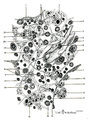 |
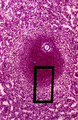
Scheme of reticular tissue in lymph node | Lymph node stroma does not contain stromal cells but instead the reticular meshwork is build by reticular cell types, macrophages and lymphoid cells and myeloid cells. Route (A) in the scheme represents morphological differentiation path from B lymphocytes to plasma cells. 1 endothelial ce... | reticular tissue; scheme; follicle; germinal center | Poja Histology Collection - Lymphatic Tissues and Organs Subset |
| 27 |
 |
Section of lymph node (human) | Stain: Silver stain (Gomori). Due to the argyrophilia the reticular fibers are black-stained. They are derived from the fibrous capsule and penetrate into the deep cortex (i.e. paracortical area) and are embedded among other fibers such as collagen type III, IV. Note the postcapillary venules (*) ... | high endothelial venule (HEV); paracortex; argyrophilia; paracortex | Poja Histology Collection - Lymphatic Tissues and Organs Subset |
| 28 |
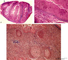 |
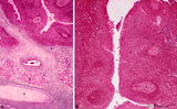
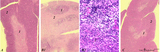
Survey and details of palatine tonsil ('lymphoepithelial tissues', 'gut-associated lymphatic tissue' or GALT) (human) | Stain: Azan. The survey in (A) shows that the palatine tonsil (localized in the lateral wall of the oropharynx) consists of crypts (1) and folds of the surface epithelium (stratified squamous) surrounded by accumulations of lymphoid cells organized in follicles (2). (B): the lining epithelium (5) i... | stratified squamous epithelium | Poja Histology Collection - Lymphatic Tissues and Organs Subset |
| 29 |
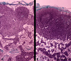 |
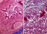
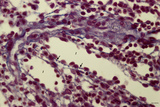
T cell depletion in lymph nodes (dog) | Stain: Trichrome (Goldner). Left and right: survey of lymph nodes. A: lymph node after treatment with anti-thymocyte-antiserum (ATS); B: normal, untreated lymph node ATS treatment results in a considerable depletion of the T cells in the paracortical area (2), while also the germinal centre (1) i... | T cell depletion; follicle; medulla; paracortex | Poja Histology Collection - Lymphatic Tissues and Organs Subset |
| 30 |
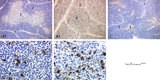 |
The effect of cyclophosphamide on the resident macrophages in thymus (rat) | Stain: Immunoperoxidase staining with diaminobenzidin (DAB) and hematoxylin counterstained on frozen section. A single injection with cyclophosphamide (CP, 70 mg/ml) induces a transient cortical involution after 4 days. The darkly stained cortex of th normal thymus (A1, A2) decreases its dark stain... | cyclophosphamide; ED1 macrophages ; immunosuppression; lymphoid tissue | Poja Histology Collection - Lymphatic Tissues and Organs Subset |
| 31 |
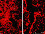 |
The effect of cyclophosphamide on B cells in spleen (rat) | Stain: Immunofluorescence of Vector red using Mark-1 antibody against B cells. A: Normal rat spleen. (1) the dark, unstained area represents the PALS (periarteriolar lymphatic sheath) filled with T cells. (2) red-stained germinal centre of a B cell follicle that is surrounded by the marginal zone (3... | cyclophosphamide; immunosuppression; immunofluorescence; B lymphocytes | Poja Histology Collection - Lymphatic Tissues and Organs Subset |
| 32 |
 |
Tubal tonsil (human) | Stain: Azan. The tubal tonsil consists of a collection of lymphoid nodules near the auditory tube opening and forms part of the Waldeyers ring of defense in the nasopharyngeal cavity. This tonsil has fewer crypts (1), and the surface is covered by one to more layered ciliated epithelium (2). The la... | tubal tonsil; nasopharynx | Poja Histology Collection - Lymphatic Tissues and Organs Subset |
| 33 |
 |
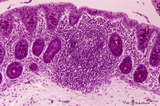
Lingual tonsil ('lymphoepithelial tissue', 'gut-associated lymphatic tissue' or GALT) (human) | Stain: Azan. A: Left side shows a secondary lymphatic follicle (1) separated by the connective tissue of the proper lamina (2) from the lining non-keratinized stratified squamous epithelium (3). The mantle zone (4) is indicated by the peripheral dense aggregations of (memory) B lymphocytes. (5) diff... | lingual tonsil; GALT; non-keratinized stratified squamous epithelium | Poja Histology Collection - Lymphatic Tissues and Organs Subset |
| 34 |
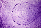 |
Border of white pulp in spleen (mouse) | Monoaminooxidase (enzyme histochemistry on frozen section) with Nitro-BT as staining substrate resulting in a blue formazan precipitate. Despite the general activity of most cells in the spleen, the border cells or so-called metallophilic cells (1) or dendritic antigen-presenting cells (APC) show th... | monoamino oxidase; ED3 antibody; follicle; white pulp | Poja Histology Collection - Lymphatic Tissues and Organs Subset |
| 35 |
 |
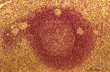
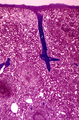
Colon ('gut-associated lymphatic tissue' or GALT) (human) | Stain: Hematoxylin-PAS. Solitary lymphatic nodule in colon (see also Digestive System: Colon) A large amount of non-encapsulated diffuse lymphatic tissue or mucosa-associated lymphatic tissue (MALT) is located in the subepithelial lamina propria of the colon and called gut-associated lymphatic tiss... | GALT; follicle; lymphoid tissue | Poja Histology Collection - Lymphatic Tissues and Organs Subset |
| 36 |
 |
Palatine tonsil ('lymphoepithelial tissues', 'gut-associated lymphatic tissue' or GALT) (human) | Stain: Azan. A low magnification in A shows the thick blue connective septum (1) with a secondary lymphatic nodule (2). (3) is a cross-section of a crypt filled with detached squamous epithelium (3a) mixed up with keratinized material (red) and a huge amount of lymphocytes (3b). A higher magnificati... | follicle; germinal center; mantle layer; stratified squamous epithelium | Poja Histology Collection - Lymphatic Tissues and Organs Subset |
| 37 |
 |
Hassall's corpuscle in thymus (human, puberty) | Stain: Hematoxylin. A larger magnification of the thymic medulla reveals the purple-stained structures in the centre of a so-called Hassall's (or thymic) corpuscle consisting of keratinized (*) epithelial cells. It is surrounded by recognizable flattened cells showing keratohyalin granules (-->). T... | thymic corpuscle (Hassalls); epithelioreticular cell (ERC); lymphoid tissue | Poja Histology Collection - Lymphatic Tissues and Organs Subset |
| 38 |
 |
Spleen with secondary lymphatic nodule (human) | Stain: Hematoxylin & eosin. The splenic follicle as part of the white pulp is arranged around the cross sectioned central artery (1).The lymphatic sheath or PALS is composed of T cells (2). The darker stained mantle zone of mainly nave B lymphocytes (3) encompasses the lighter stained germinal centr... | white pulp; PALS; marginal zone; mantle layer | Poja Histology Collection - Lymphatic Tissues and Organs Subset |
| 39 |
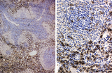 |
Immunohistochemistry of ED1-positive subset of macrophages in spleen (rat) | Stain: Immunoperoxidase staining using diaminobenzidin (DAB)/ hematoxylin counterstained on frozen section with the anti-macrophage antibody ED1. Most of the labeled (brown) macrophages are found in the red pulp (2) up to the marginal zone border (B). The PALS area (1) contains sparsely spread ED1 ... | ED1 ; macrophages; immunohistochemistry; marginal zone | Poja Histology Collection - Lymphatic Tissues and Organs Subset |
| 40 |
 |
Spleen with secondary lymphatic nodule (human) | Stain: Azan. The white pulp of this perfused spleen consists of: at (1) a cross-section of the central artery, (2) tangential cut mantle zone and (3) the marginal zone. The red pulp contains empty venous sinusoids (4) and the perilymphoid zone (3a) is the zone of red pulp immediately surrounding ... | white pulp; germinal center; follicle; white pulp | Poja Histology Collection - Lymphatic Tissues and Organs Subset |
| 41 |
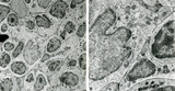 |
Part of lymphatic nodule in spleen (rat) | Electron microscopy. The left image (A) reveals part of a white pulp area stuffed with a dendritic cell (1) between a majority of different types of lymphocytes (2, 3). The right image shows a larger magnification of the same area with the dendritic cell (1) sandwiched in between the enclosing lymp... | dendritic cell; electron microscopy; white pulp | Poja Histology Collection - Lymphatic Tissues and Organs Subset |
| 42 |
 |
Thymus after cyclophosphamide treatment (rat) | Stain: Hematoxylin & eosin. A single injection with cyclophosphamide (CP, 4 70 mg/ml) induces a transient cortical involution, i.e. inhibition of the cell proliferation and maturation. A: Normal thymus with medulla (1) and cortex (2). B1: Inversion of thymic cortex and medulla 4 to 8 days after CP ... | cyclophosphamide; immunosuppression; involution; lymphoid tissue | Poja Histology Collection - Lymphatic Tissues and Organs Subset |
| 43 |
 |
Lymphoblast in splenic white pulp (rat, human) | Electron microscopy (A, rat) and Methyl green (B, human). Upon antigenic stimulation the lymphocytes in the germinal centre proliferate and generate activated B cells or lymphoblasts which seed towards marginal zone and red pulp while differentiating. Due to the increased number of lymphoblasts, ret... | electron microscopy; lymphoblast | Poja Histology Collection - Lymphatic Tissues and Organs Subset |
| 44 |
 |
Phagocytosis in small splenic blood vessel (mouse) | Electron microscopy. Stain: Peroxidase reaction with diaminobenzidin staining. A diversity of red blood cells is black-stained due to the staining of hemoglobin by oxidized benzidin. Circulating lymphocytes (2) in the lumen (*) remain unstained. An oblong monocyte (1) developing into a macrophage h... | electron microscopy; phagocytosis | Poja Histology Collection - Lymphatic Tissues and Organs Subset |
| 45 |
 |
Survey of spleen (human) | Stain: Azan. The spleen is covered by a capsule (1) of dense connective tissue and elastic fibers. The capsule continues into the spleen as trabeculae (2) carrying blood vessels and nerve fibers. As arteries leave the trabeculae it becomes invested by a sheath of T cells forming a PALS (3) or periar... | white pulp; sinusoid; red pulp; PALS | Poja Histology Collection - Lymphatic Tissues and Organs Subset |
| 46 |
 |
: Lymph node (human) | Stain: Azan. Specialized venules (1) or so-called high endothelial venules (HEV) are here located in the paracortical area (4) close to the lymphatic follicle (2+3). The HEVs are lined by cuboidal or columnar endothelial cells that possess specific homing receptors for antigen-primed B- and T ly... | paracortex; high endothelial venule (HEV); germinal center | Poja Histology Collection - Lymphatic Tissues and Organs Subset |
| 47 |
 |
Red pulp of spleen with perfused venous sinusoids (human) | Stain: Azan. Cross-sectioned venous sinusoids with splenic cord (1). The wall of a sinusoid is composed of elongated rod-like endothelial cells that are orientated parallel to each other in the long axis of the sinusoid. There is a discontinuous pale-stained basement membrane (difficult to observe i... | sinusoid ; red pulp; splenic cord; central arteriole | Poja Histology Collection - Lymphatic Tissues and Organs Subset |
| 48 |
 |
Venous circulation pattern in perfused spleen (human) | Stain: Azan. The composed picture shows part of the splenic circulation system at several enlargements (inset, A, B). The open venous sinusoids (1) drain via short pulp veins into thin-walled trabecular veins (2), subsequently into thick-walled trabecular veins (4). The trabeculae originate from the... | splenic circulation; trabecular veins; sinusoid ; PALS | Poja Histology Collection - Lymphatic Tissues and Organs Subset |
| 49 |
 |
Thymus medulla (rat, young adult) | Electron microscopy. Epithelioreticular cells of the medulla (1) close to each other. The electron-light cytoplasm contains many small vesicles (1, Golgi area) as well as cross-sections of vacuoles (2) with small finger-like cytoplasmic extrusions in the lumen. Electron-dense lysosomal structures (3... | medullar epithelioreticular cell ; thymus medulla; lymphoid tissue | Poja Histology Collection - Lymphatic Tissues and Organs Subset |
| 50 |
 |
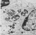
Thymus medulla (rat, young adult) | Electron microscopy. Surrounded by thymocytes (3) a medullary macrophage with an electron-light nucleus (1). The cytoplasm contains many electron-dense lysosomes of varying sizes and forms (2). | medullar macrophage; lymphoid tissue | Poja Histology Collection - Lymphatic Tissues and Organs Subset |