The Health Education Assets Library (HEAL) is a collection of over 22,000 freely available digital materials for health sciences education. The collection is now housed at the University of Utah J. Willard Marriott Digital Library.
TO
Filters: Collection: "ehsl_heal"
| Title | Description | Subject | Collection | ||
|---|---|---|---|---|---|
| 26 |
 |
Elastin in alveolar tip in lung tissue (human, adult) | Immuno-electron microscopy (embedded in Lowicryl HM 20). The amorph elastin (E) appears pale after embedding in this type of resin, but is antibody-labeled with electron-dense gold particles of 10 nm. (Col) indicate bundles of collagen fibers which are intermingled with swollen cytoplasmic extension... | Alveolar cell type I; Myofibroblast; Immuno-electron microscopy; Immuno-staining | Poja Histology Collection - Respiratory System Subset |
| 27 |
 |
Elastin in alveolus of lung tissue (human, adult) | Immuno-electron microscopy (embedded in Lowicryl HM 20). Elastin (E) is labeled with electron-dense gold particles of 10 nm. Note that the amorph elastin appears pale after embedding in this type of resin. (A) indicates alveolar space, (F) unlabelled myofibrolast, (C) collagen fibers, and (1) the th... | Alveolar cells type I; Myofibroblasts; Immuno-electron microscopy; Immuno-staining | Poja Histology Collection - Respiratory System Subset |
| 28 |
 |
Elastin in alveolus of lung tissue (human, adult) | Immuno-electron microscopy (embedded in Lowicryl HM 20). The amorph elastin (E) appears pale after embedding in this type of resin, but is antibody-labeled with electron-dense gold particles of 10 nm. (C) indicate bundles of collagen fibers (unlabeled) which are intermingled with swollen cytoplasmic... | Immuno-staining; Immuno-electron microscopy | Poja Histology Collection - Respiratory System Subset |
| 29 |
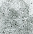 |
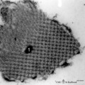
Elastin in lung arteriole (human, adult) | Immuno-electron microscopy (embedded in Lowicryl HM 20). The alveolar septa contain a muscular pulmonary arteriole (lumen = X) with unlabelled endothelial cells (1). The elastic membranes in the arteriolar walls consist of more or less continuous bands of amorphous elastic lumps (E) antibody-labeled... | Alveolar septum; Immuno-electron microscopy; Immuno-staining | Poja Histology Collection - Respiratory System Subset |
| 30 |
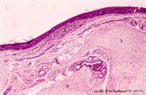 |
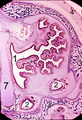
Epiglottis (human) | Stain: Hematoxylin and eosin. Stratified squamous epithelium (1) at the laryngeal side (top). Lymphocyte accumulation (2) in the lamina propria. Below elastic cartilage (3) and seromucous laryngeal glands (4) with draining ducts. | Squamous epithelium; Laryngeal glands | Poja Histology Collection - Respiratory System Subset |
| 31 |
 |
Epithelial lining of bronchiolus in the lung (mammalia) | Scheme electron microscopy. Two major cell types line a bronchiolus, a more cuboidal shaped ciliated one (1) that also exhibits microvilli with many organelles (including electron-dense lysosomal structures). The non-ciliated cells (2, Clara cells) appeared taller and dome-shaped and protrude into t... | Bronchiolus; Ciliated epithelium; Clara cells | Poja Histology Collection - Respiratory System Subset |
| 32 |
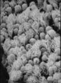 |
Epithelial lining of bronchiolus in the lung (rat) | Scanning electron microscopy. Bushes of cilia indicate the presence of ciliated cells (1). Clustered non-ciliated cells (2, Clara cells) are dome-shaped with stubby microvilli at the surface, they protrude into the lumen to the tips of the cilia. | Bronchiolus ; Ciliated epithelium ; Clara cells | Poja Histology Collection - Respiratory System Subset |
| 33 |
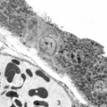 |
Epithelial lining of respiratory bronchiolus in the lung (gerbil) | Electron microscopy (low magnification). Two major cell types line a bronchiolus: a cuboidal-shaped ciliated one that exhibits microvilli with many organelles (1); the non-ciliated cells (2, Clara cells) are taller and are well provided with numerous electron-grey and smaller electron-dense secretor... | Respiratory bronchiolus; Ciliated epithelium; Cuboidal epithelium; Clara cells; Secretory granules | Poja Histology Collection - Respiratory System Subset |
| 34 |
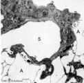 |
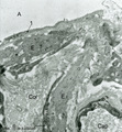
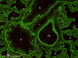
Epithelial lining of respiratory bronchiolus in the lung (rat) | Electron microscopy (low magnification). The epithelial lining of the respiratory bronchiolus contains ciliated cells (1) and Clara cells (2) with electron-dense secretory granules. At (4) interstitium with elastin, collagen and myofibroblasts; (5) shows a small pulmonary arteriole. At arrow (→) a... | Respiratory bronchiolus; Ciliated epithelium; Clara cells; Secretory granules; Pneumocytes; Alveolar cells; Interstitium | Poja Histology Collection - Respiratory System Subset |
| 35 |
 |
Epithelial lining of terminal bronchiolus (golden hamster) | Electron microscopy (low magnification). Two major epithelial cell types line the bronchiolus: (1) the ciliated cells with many organelles, and (2) the non-ciliated cells or Clara cells well provided with organelles, especially agranular endoplasmic reticulum and depending on their activity, numerou... | Terminal bronchiolus; Ciliated epithelium ; Clara cells; Neuro-endocrine cells; Pneumocytes; Alveolar cell types; Interstitium | Poja Histology Collection - Respiratory System Subset |
| 36 |
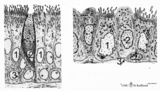 |
Epithelial lining of trachea and bronchiolus (mammals) | Left side trachea: Columnar ciliated cells (1) with up to 200 motile cilia with an organelle-rich apex and in the middle a goblet cell (2) with accumulation of mucus secretion granules in the apex. Note that all epithelial cells (i.e., junctions) contact the basal lamina. Four basal cells (3) do not... | Bronchiolus; Pseudostratified epithelium; Ciliated epithelium; Clara cells; Basal cells; Neuro-endocrine cells | Poja Histology Collection - Respiratory System Subset |
| 37 |
 |
Epithelium of respiratory bronchiolus (detail, human, high magnification) | Stain: Azan. An irregular lining of low columnar-cuboidal cells (1) and thin alveolar epithelium (2) are present. Note cuboidal Clara cells (↓). At (3) small patches of smooth muscles and at (4) macrophages with phagocytized carbon particles (black dots). Small pulmonary artery (5), lymph capillar... | Respiratory bronchiolus; Cuboidal epithelium; Alveolar epithelium; Lymph capillary | Poja Histology Collection - Respiratory System Subset |
| 38 |
 |
Epithelium of trachea (golden hamster) | Electron microscopy. Columnar ciliated cells (1) with cilia and organelle-rich apex; a goblet cell (2a) with accumulation of mucous secretion granules supranuclearly. Another goblet cell (2b) appears deprived; one basal cell (3) does not extend to the free surface. Light-grey aggregations of elastin... | Respiratory epithelium ; Pseudostratified epithelium; Basal cells | Poja Histology Collection - Respiratory System Subset |
| 39 |
 |
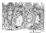
Epithelium of trachea (mammals, common respiratory epithelium) | Scheme electron microscopy. Columnar ciliated cells (1) with up to 200 motile cilia with an organelle-rich apex and in the middle a goblet cell (2) with accumulation of mucus secretion granules in the apex. Note that all epithelial cells (i.e., junctions) contact the basal lamina. Four basal cells (... | Respiratory epithelium ; Pseudostratified epithelium; Basal cells | Poja Histology Collection - Respiratory System Subset |
| 40 |
 |
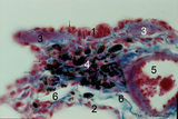
Fibrillin in the alveoli in lung tissue (human, adult) | Stain: imunoperoxidase staining with anti-fibrillin antibodies and diaminobenzidin reaction (frozen section). Fibrillin is one of the elastin-associated microfibrillar proteins that wraps the elastin protein core and is localized in normal lung structures such as alveolar septa and tips. The immuno-... | Alveolar tips; Elastin-associated proteins; Fibrillin | Poja Histology Collection - Respiratory System Subset |
| 41 |
 |
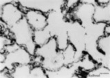
Fibrillin in the alveoli of lung emphysema (human, adult) | Stain: antifibrillin antibody immunoperoxidase staining with diaminobenzidin reaction. Fibrillin is one of the elastin-associated microfibrillar proteins, and marks therefore also the presence of elastin in lung tissue. In centrilobular emphysema (e.g., in lungs of smokers) the lesions are more com... | Alveolar septa; Elastin-associated proteins; Fibrillin | Poja Histology Collection - Respiratory System Subset |
| 42 |
 |
Fibrillin in the alveoli of lung emphysema (human, adult) | Stain: antifibrillin antibody immunoperoxidase staining with diaminobenzidin reaction. Fibrillin is one of the elastin-associated microfibrillar proteins, and marks therefore the presence of elastin in lung tissue. In centrilobular emphysema (e.g., in lungs of smokers) the lesions are more common an... | Alveolar tips; Elastin-associated proteins; Fibrillin | Poja Histology Collection - Respiratory System Subset |
| 43 |
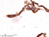 |
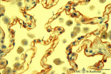
Fibrillin in the alveoli of the lung (human, adult) | Stain: antifibrillin antibody immunoperoxidase staining with diaminobenzidin reaction. Fibrillin is one of the elastin-associated microfibrillar proteins. Here fibrillin (and elastin) is localized as brown threads in the alveolar septa and alveolar tips (1). The elastin is visible as white threads (... | Alveolar tips; Elastin-associated proteins; Fibrillin | Poja Histology Collection - Respiratory System Subset |
| 44 |
 |
Free surfactant (tubular myelin) in alveolar space of the lung (rat) | Electron microscopy. After fixation the extracellular lining of surfactant (phosphatidylcholine, phosphoglycerol, cholesterol and proteins) will often be present as free packed lamellae in the alveolar space. This so-called tubular myelin (partly cross-sectioned), (1) is observed as stacks of lipid ... | Pneumocyte II; Tubular myelin | Poja Histology Collection - Respiratory System Subset |
| 45 |
 |
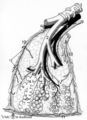
Frontal section of head (pig, fetus) | Stain: Azan. At the upper half the nasal septum (7) is a lightly stained plate (cartilago septi nasi). Developing conchae with supporting hyaline cartilage scaffolds (*) (light-stained) are present in the nasal chamber (1) and known as inferior (lowest), middle and superior turbinate bones. The whol... | Conchae nasales; Trabecular bone | Poja Histology Collection - Respiratory System Subset |
| 46 |
 |
Gross scheme of a lung lobule and its vascularization (human) | Scheme of branching of intrapulmonary bronchus. A small intrapulmonal bronchus with cartilage rings (1) divides into bronchioli (2), at (3) the terminal bronchioli that continues into respiratory bronchioli (4) followed by alveolar ducts (5) and at (6) the alveoli. A respiratory bronchiolus with alv... | Intrapulmonary bronchus; Bronchioli; Alveolar ducts; Lymphatic plexus; Visceral pleura | Poja Histology Collection - Respiratory System Subset |
| 47 |
 |
Heparan sulfates in the terminal sac period of the lung (mouse, fetus) | Stain: fluorescence microscopy with anti-heparan sulphate antibody (a phage-display antibody, HS4E4). Heparan sulfates are linear polysaccharides that belong to the group of the glycosaminoglycans (GAGs). These GAGs are found associated with basement membranes as shown here. The sulphated saccharide... | Bronchioli; Air spaces; Terminal sac period | Poja Histology Collection - Respiratory System Subset |
| 48 |
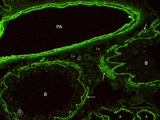 |
Heparan sulfates in the terminal sac-period of the lung (mouse, fetus) | Stain: fluorescence microscopy with anti-heparan sulphate antibody (a phage-display antibody, LKiv69). Heparan sulfates are linear polysaccharides that belongs to the group of the glycosaminoglycans (GAGs). The sulphated saccharide domains provide numerous docking sites for protein ligands. These GA... | Bronchioli; Air spaces; Terminal sac period | Poja Histology Collection - Respiratory System Subset |
| 49 |
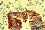 |
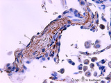
Keratin 7 in the bronchial epithelium of the lung (human, adult) | Stain: anti-keratin 7 antibody (Pan-Ck 7) immunoperoxidase staining (with aminoethylcarbazole (AEC) substrate). The epithelium of the bronchus (1) stains dark brown-red after the reaction with AEC indicating a positive reaction for cytokeratin 7. This antibody can be used in the study of development... | Bronchiolus; Immunoperoxidase; Immuno-reaction | Poja Histology Collection - Respiratory System Subset |
| 50 |
 |
Keratin 7 in the lining alveolar epithelium of the lung (human, adult) | Stain: anti-keratin 7 antibody (Pan-Ck 7) immunoperoxidase staining (with aminoethylcarbazole (AEC) substrate). Both alveolar epithelial cell types I (1) and II (2) stain dark brown-red after the reaction with AEC indicating a positive reaction for cytokeratin 7. Note the white (non-stained) capilla... | Pneumocyte I; Pneumocyte II; Imunoperoxidase; Immuno-reaction | Poja Histology Collection - Respiratory System Subset |