The Health Education Assets Library (HEAL) is a collection of over 22,000 freely available digital materials for health sciences education. The collection is now housed at the University of Utah J. Willard Marriott Digital Library.
TO
Filters: Collection: "ehsl_heal"
| Title | Description | Subject | Collection | ||
|---|---|---|---|---|---|
| 26 |
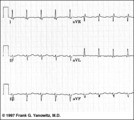 |
Frontal plane QRS axis = -45 degrees | Frontal plane QRS axis = -45 degrees | Knowledge Weavers ECG | |
| 27 |
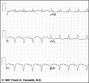 |
Frontal plane QRS axis = -75 degrees | Frontal plane QRS axis = -75 degrees | Knowledge Weavers ECG | |
| 28 |
 |
Neuropile, anterior horn cells, myelinated axons | Neuropile, anterior horn cells, myelinated axons. Thin plastic section of spinal cord. Photograph. Multimedia. | Axons; Spinal cord; Brain; Central nervous system; Anatomy | Slice of Life |
| 29 |
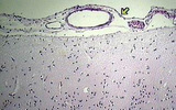 |
Arachnoid with pial vessels and pia and cerebral cortex | Arachnoid with pial vessels and pia and cerebral cortex. Photograph. Multimedia. | Arachnoid; Cerebral veins; Cerebral cortex; Brain; Central nervous system; Anatomy | Slice of Life |
| 30 |
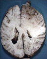 |
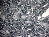
Glioblastoma multiforme, frontal lobe | Glioblastoma multiforme, frontal lobe. Case PM86-83, resection of right pole-spread to left. Horizontal plane. Photograph. Multimedia. | Glioblastoma; Brain; Frontal lobe; Central nervous system; Anatomy | Slice of Life |
| 31 |
 |
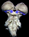
Hypothalamus and Relationship to Posterior Pituitary (Labeled) | Hypothalamus. Posterior pituitary. | Supraoptic Hypothalamic Nucleus; Optic Chiasma; Pituitary Stalk; Adenohypophysis; Pars Tuberalis; Pars Intermedia; Pars Distalis; Neurohypophysis | Royal College of Surgeons in Ireland Illustrations |
| 32 |
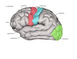 |
Sensory Cortex, Visual Cortex, Auditory Cortex and Motor Cortex - Lateral (Labeled) | Sensory, visual, auditory, and motor cortices. | Central Sulcus; Sensory Area; Lateral Fissure; Auditory Area | Royal College of Surgeons in Ireland Illustrations |
| 33 |
 |
Superior colliculus, brachium and medial geniculate | Superior colliculus, brachium and medial geniculate. Graphic overlay on dorsal surface of brain stem. Photograph. Multimedia. | Superior colliculus; Geniculate bodies; Brain stem; Brain; Central nervous system; Anatomy | Slice of Life |
| 34 |
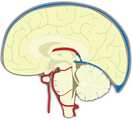 |
Brain - Midsagittal Schematic Section with Major Arteries and Venous Sinuses | Schematic section of the brain showing major arteries and venous sinuses. | Foramen of Monro; Splenium of Corpus Callosum; Genu of Corpus Callosum; Posterior Calcarine Sulcus | Royal College of Surgeons in Ireland Illustrations |
| 35 |
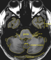 |
MRI Atlas: Brain (Axial) - Scan 2 - Labeled | Anatomical structures of the brain are identified in this scan with labeled outline from the UCLA Interactive Neurosciences MRI Atlas. | UCLA Interactive Neuroscience | |
| 36 |
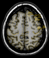 |
MRI Atlas: Brain (Axial) - Scan 8 - Labeled | Anatomical structures of the brain are identified in this scan with labeled outline from the UCLA Interactive Neurosciences MRI Atlas. | UCLA Interactive Neuroscience | |
| 37 |
 |
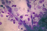
Bone Marrow with Megaloblastic Erythropoiesis | BM with moderate megaloblastic erythropoiesis | Ea1400 | Albert Einstein College of Medicine Gallery of Hematology Images |
| 38 |
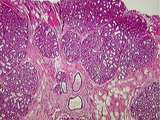 |
Breast | This active, lactating breast contains a large number of lobules of tightly packed alveoli. The connective tissue of the interlobular elements is highly compressed. Note the lactating glands, the excretory duct and some fat. UCLA Histology Collection. | Breast; lactating breast | UCLA Histology |
| 39 |
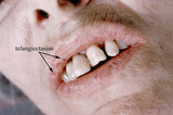 |
Hereditary Hemorrhagic Telangectasia | patient with Hereditary Hemorrhagic Telangiectasia | Da090T; Microcytic Anemia; HHT | Albert Einstein College of Medicine Gallery of Hematology Images |
| 40 |
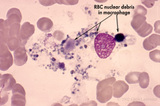 |
Intramedullary Hemolysis from Histiocytes Phagocytising Megaloblasts | intramedullary hemolysis from histiocytes phagocytising megaloblasts | Ea15aT | Albert Einstein College of Medicine Gallery of Hematology Images |
| 41 |
 |
Normal Vessel in Bone Marrow | normal vessel in bone marrow (400x) | Kg06a0 | Albert Einstein College of Medicine Gallery of Hematology Images |
| 42 |
 |
Plasmodium malariae with Intracellular Ring Forms | P malariae with intracellular ring forms | Vf0900 | Albert Einstein College of Medicine Gallery of Hematology Images |
| 43 |
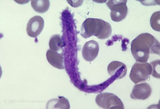 |
Platelet in Myelofibrosis | giant, worm-like platelet in myelofibrosis | Jd0500; MPD | Albert Einstein College of Medicine Gallery of Hematology Images |
| 44 |
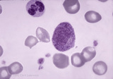 |
Promyelocyte | mature promyelocyte | Ba15a0; White Blood Cells; WBC | Albert Einstein College of Medicine Gallery of Hematology Images |
| 45 |
 |
Skin Tumor | Tumors of various sorts can be produced by anything that grows within the dermis.This demonstrates that the tumor was within the skin, and moves freely with the skin. | Knowledge Weavers Dermatology | |
| 46 |
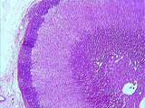 |
Adrenal | The classical organization of the adrenal gland with a thick capsule and the cortex composed of three layers: the outer zona glomerulosa, the middle zona fasciculata and the inner zona reticularis. Finally, in the center of the gland is the adrenal medulla. UCLA Histology Collection. | Adrenal | UCLA Histology |
| 47 |
|
Adrenal | The classical organization of the adrenal gland with a thick capsule and the cortex composed of three layers: the outer zona glomerulosa, the middle zona fasciculata and the inner zona reticularis. Finally, in the center of the gland is the adrenal medulla. UCLA Histology Collection. | Adrenal | UCLA Histology |
| 48 |
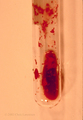 |
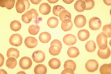
Agglutination of Red Blood Cells | agglutination of RBCs at room temperature in CBC tube | Ec0400; Cold Agglutinin Disease; CAD | Albert Einstein College of Medicine Gallery of Hematology Images |
| 49 |
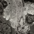 |
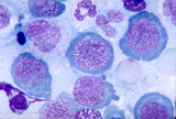
Dendritic cells in spleen (mouse) | Electron microscopy. The interdigitations (*) of the left dendritic cells (antigen-presenting cell or APC) (1 and 2a) are clearly shown. The right (2b) is a neighbour dendritic cell sectioned at the level of the Golgi area. (3) lymphocyte and (4) reticular cell. | electron microscopy; dendritic cell | Poja Histology Collection - Lymphatic Tissues and Organs Subset |
| 50 |
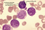 |
Immature Eosinophile with both Eosin- and Basophilic Granules | immature eosinophile with both eosin- & basophilic granules in CML blast crisis | Ja06aT; WBC | Albert Einstein College of Medicine Gallery of Hematology Images |