The Health Education Assets Library (HEAL) is a collection of over 22,000 freely available digital materials for health sciences education. The collection is now housed at the University of Utah J. Willard Marriott Digital Library.
TO
Filters: Collection: "ehsl_heal"
| Title | Description | Subject | Collection | ||
|---|---|---|---|---|---|
| 401 |
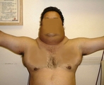 |
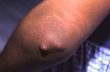
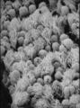
Multiple Systemic Lipomatosis | Male patient with multiple systemic lipomatosis. | Multiple Systemic Lipomatosis | HEAL Reviewed Collection |
| 402 |
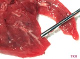 |
Wet hepatisized lung at autopsy | Wet hepatisized lung at autopsy of infant dying of Type II Meconium Aspiration Syndrome (MAS). | Type II MAS | Harris Pediatric Image Collection |
| 403 |
 |
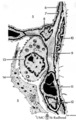
Diastomyelia | This image depicts diastomyelia. | Split Cord; Myeloschisis; Hemi Cord; Congenital Scoliosis; Tethered Cord; Spinal Cord Myelodysplasia | HEAL Reviewed Collection |
| 404 |
 |
Midbrain | Midbrain. Superior colliculus, red nucleus, III nucleus. Transverse plane. Photograph. Multimedia. | Mesencephalon; Superior colliculus; Red nucleu; Cranial nerves; Oculomotor nerve; Central nervous system; Anatomy | Slice of Life |
| 405 |
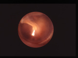 |
Tympanic Membrane | Normal tympanic membrane | Ear Drum | HEAL Reviewed Collection |
| 406 |
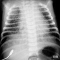 |
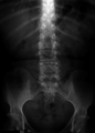
Chest X-ray of (MAS) infant with severe Type I Meconium Aspiration Syndrome prior to respiratory failure | Chest X-ray of baby with severe Type I Meconium Aspiration Syndrome (MAS) just prior to respiratory failure (RF) requiring mechanical ventilation - noting patchy infiltrates or areas of atelectasis scattered throughout markedly over-expanded lungs (predominantly on the right) due to air-trapping. | Air Trapping; Respiratory Failure; Infiltrates | Harris Pediatric Image Collection |
| 407 |
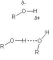 |
General Description of Hydrogen Bonding in Alcohols | This image depicts the basic interaction between the oxygen in one alcohol with the hydrogen of an adjccent alcohol. The oxygen is shown with a partial negative charge (as shown by the negative lowercase delta), and the hydrogen of the adjacent alcohol has the corresponding partial postive charge (p... | HEAL Open Review Collection | |
| 408 |
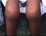 |
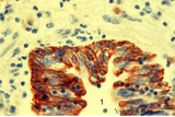
Arthritis of Both Knees | Arthritis of large joints occurs with polyarticular or pauciarticular disease, while small joint involvement is typically seen in polyarticular disease only. | Polyarticular Juvenile Rheumatoid Arthritis; Pauciarticular Juvenile Rheumatoid Arthritis | HEAL Reviewed Collection |
| 409 |
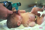 |
Manual ventilation during successful delivery room resuscitation | Manual ventilation during successful delivery room resuscitation of a meconium-stained, initially depressed, postmature newborn infant male. | manual ventilation | Harris Pediatric Image Collection |
| 410 |
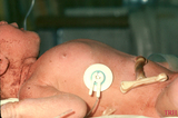 |
Increased AP-diameter of chest in MAS infant due to air-trapping | Increased AP-diameter of chest in infant with Meconium Aspiration Syndrome (MAS) due to air-trapping. | Air Trapping; Anterior-Posterior Chest Diameter | Harris Pediatric Image Collection |
| 411 |
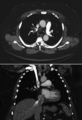 |
Pulmonary Embolus | This image contains two scans of the same Pulmonary Embolus found in a patient. | Pulmonary Infarction; Westermark Sign; Intraluminal Filling Defects, Subsegmental; Hampton Hump; Pleural Based Density | HEAL Reviewed Collection |
| 412 |
 |
Suctioning of the mouth in MAS infant before delivery of the body | Suctioning of the mouth of infant with Meconium Aspiration Syndome (MAS) before delivery of the body. | Mouth Suctioning | Harris Pediatric Image Collection |
| 413 |
 |
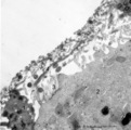
Subcutaneous Nodules | Rheumatoid nodules occur more commonly in rheumatoid factor positive children with advanced disease. | HEAL Reviewed Collection | |
| 414 |
 |
Lack of classic inflammation in subject with severe asthma | Endobronchial biopsy from severe asthmatic with minimal inflammation, but abundant mucus and airway edema. | HEAL Reviewed Collection | |
| 415 |
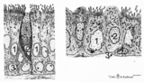 |
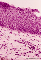
Epithelial lining of trachea and bronchiolus (mammals) | Left side trachea: Columnar ciliated cells (1) with up to 200 motile cilia with an organelle-rich apex and in the middle a goblet cell (2) with accumulation of mucus secretion granules in the apex. Note that all epithelial cells (i.e., junctions) contact the basal lamina. Four basal cells (3) do not... | Bronchiolus; Pseudostratified epithelium; Ciliated epithelium; Clara cells; Basal cells; Neuro-endocrine cells | Poja Histology Collection - Respiratory System Subset |
| 416 |
 |
Air-blood barrier in the lung (mammals) | Scheme electron microscopy. (1, ↓) Represents type I pneumocytes lining alveolar spaces (A). Cell (2) represents a free alveolar macrophage. The type II pneumocyte (3) is adherent to type I pneumocyte extensions (note junctional connection), and contains multilamellar bodies (surfactant). A myofib... | Pneumocyte type I ; Pneumocyte type II | Poja Histology Collection - Respiratory System Subset |
| 417 |
 |
Neuroepithelial body in terminal bronchiolus (golden hamster) | Electron microscopy. Three epithelial cells, as part of the N(euro) E(pithelial) B(ody), contribute to a cluster of neuroendocrine cells. They belong to the Amine Precursor Uptake and Decarboxylation cell system so-called APUD cells. The dense-core granules (↓) contain among others dopamine or ser... | Terminal bronchiolus ; Neuro-endocrine cells | Poja Histology Collection - Respiratory System Subset |
| 418 |
 |
Keratin 7 in the bronchial epithelium of the lung (human, adult) | Stain: anti-keratin 7 antibody (Pan-Ck 7) immunoperoxidase staining (with aminoethylcarbazole (AEC) substrate). The epithelium of the bronchus (1) stains dark brown-red after the reaction with AEC indicating a positive reaction for cytokeratin 7. This antibody can be used in the study of development... | Bronchiolus; Immunoperoxidase; Immuno-reaction | Poja Histology Collection - Respiratory System Subset |
| 419 |
 |
Surface of olfactory epithelium (rat) | Electron microscopy. Bottom left shows an olfactory bulb (1, vesicle) with cross-sectioned basal bodies. Right side part of the apex of a supporting cell (2) with microvilli (3). Parallel to the surface of the epithelium one long olfactory cilium (4) and several other cross-sectioned ones are detect... | Olfactory epithelium; Olfactory vesicle | Poja Histology Collection - Respiratory System Subset |
| 420 |
 |
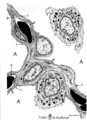
Alveolar cells in the lung (mammals) | Scheme electron microscopy. (5) alveolar space; (6) type I Pneumocyte; (7) basal lamina; (8) myofibroblast; (9) collagen and elastin fibers; (10) mesothelial cell of the visceral pleura; (11) capillary with erythrocyte; (12) endothelial cell lining the capillary; (13) type II pneum... | Pneumocyte type I ; Pneumocyte type I I | Poja Histology Collection - Respiratory System Subset |
| 421 |
 |
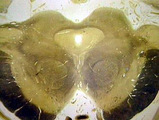
Scheme of the epiglottis (human, adult) | The laryngeal side of the epiglottis (1) is covered with respiratory epithelium, while the lingual side (2) is similar to the oral cavity epithelium (squamous type). The transitional zone to respiratory epithelium is marked by (3). The scaffold consists of elastic cartilage (4). Seromucous laryngea... | Laryngeal glands; Oral cavity | Poja Histology Collection - Respiratory System Subset |
| 422 |
 |
Epithelial lining of bronchiolus in the lung (rat) | Scanning electron microscopy. Bushes of cilia indicate the presence of ciliated cells (1). Clustered non-ciliated cells (2, Clara cells) are dome-shaped with stubby microvilli at the surface, they protrude into the lumen to the tips of the cilia. | Bronchiolus ; Ciliated epithelium ; Clara cells | Poja Histology Collection - Respiratory System Subset |
| 423 |
 |
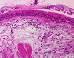
Transitional region of epiglottis (human, high magnification) | Stain: Hematoxylin and eosin. Transition of squamous epithelium into pseudostratified epithelium. Note diapedesis of lymphocytes (darker stained rounded cells) through the epithelial barrier (→). The proper lamina presents some lymphocyte infiltration. | Diapedesis | Poja Histology Collection - Respiratory System Subset |
| 424 |
 |
Keratin 7 in the lining alveolar epithelium of the lung (human, adult) | Stain: anti-keratin 7 antibody (Pan-Ck 7) immunoperoxidase staining (with aminoethylcarbazole (AEC) substrate). Both alveolar epithelial cell types I (1) and II (2) stain dark brown-red after the reaction with AEC indicating a positive reaction for cytokeratin 7. Note the white (non-stained) capilla... | Pneumocyte I; Pneumocyte II; Imunoperoxidase; Immuno-reaction | Poja Histology Collection - Respiratory System Subset |
| 425 |
 |
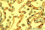
Arbor bronchialis, the bronchial tree (human, adult) | Resin corrosion cast of the lower trachea and bronchial tree (posterior aspect). The lobar and segmental bronchi and their main branches are coloured, different colours indicate areas supplied by different segmental bronchi. White-coloured lower trachea (Tr) divides into two principal bronchi (Bp)... | Segmental bronchi ; Macroscopy | Poja Histology Collection - Respiratory System Subset |