The Health Education Assets Library (HEAL) is a collection of over 22,000 freely available digital materials for health sciences education. The collection is now housed at the University of Utah J. Willard Marriott Digital Library.
TO
Filters: Collection: ehsl_heal
| Title | Description | Subject | Collection | ||
|---|---|---|---|---|---|
| 376 |
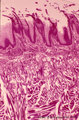 |
Papillae filliformes of the tongue (dorsal side, human) | Stain: Heidenhain light bordeaux. Threadlike keratinized extensions of the stratified epithelium. Primary connective tissue papillae with 2 to 3 secondary papillae. The skeletal muscle fibers are arranged in three directions. | oral cavity; filiform papillae | Poja Histology Collection - Oral Cavity Subset |
| 377 |
 |
Parotid gland (human) | Stain: Mallory trichrome. Survey: at the left bottom a large interlobular duct (in lumen remnants of secretion products) within a septum of dense connective tissue. At the top thinner (blue) septum, a thick (red-bluish) septum at the left. In the center three (intralobular) striated ducts between th... | oral cavity; serous gland | Poja Histology Collection - Oral Cavity Subset |
| 378 |
 |
Parotid gland (human) | Stain: Azan. The parotid gland: in most species the gland is composed entirely of serous acini. At the right a small (intralobular) striated duct; centrally one large interlobular duct with blood vessels. Scattered a few (white) fat cells. | oral cavity; serous gland | Poja Histology Collection - Oral Cavity Subset |
| 379 |
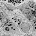 |
Parotid gland (rat) | Electronmicroscopy. Part of a serous acinus with characteristic secretion granules supranuclearly. Note different densities of the granules without any signs of fusion. | oral cavity; serous gland | Poja Histology Collection - Oral Cavity Subset |
| 380 |
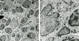 |
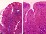
Part of lymphatic nodule in spleen (rat) | Electron microscopy. The left image (A) reveals part of a white pulp area stuffed with a dendritic cell (1) between a majority of different types of lymphocytes (2, 3). The right image shows a larger magnification of the same area with the dendritic cell (1) sandwiched in between the enclosing lymp... | dendritic cell; electron microscopy; white pulp | Poja Histology Collection - Lymphatic Tissues and Organs Subset |
| 381 |
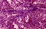
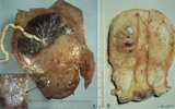 |
Partial hydatidiform mole and invasive mole (human) | (A) Macroscopy: The partial mole (1) occupies a large part of the placenta and is distinct from the normal chorionic plate where the umbilical cord (2) inserts eccentrically with branches of the umbilical vessels. The inset shows a circumscript area with swollen transparent grape-like vesicles (chor... | partial hydatidiform mole ; chorioadenoma | Poja Histology Collection - Placenta |
| 382 |
 |
Penicillar arterioles in spleen (human) | Stain: Azan. The central or follicular artery in the follicle of the spleen splits into many arterioles. These arterioles spread as so-called penicillar arterioles (1) shown here. They are still surrounded by a very thin perilymphatic sheath (PALS) that disappears as the arteries in a brush-like pat... | penicillar arterioles; red pulp | Poja Histology Collection - Lymphatic Tissues and Organs Subset |
| 383 |
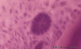 |
Periodontal ligament with epithelial rest of Malassez - longitudinal section of root of tooth, higher magnification; human, adult | Stain: Hematoxylin and eosin. Centrally within the connective tissue of the periodontal ligament a distinct darker stained epithelial islet with nuclei is present (epithelial rest of Malassez as a remnant of Hertwig's epithelial root sheath; in adults it might produce dental cyst). At the right side... | oral cavity; cementoblasts; epithelial rest of Malassez; cementoid | Poja Histology Collection - Oral Cavity Subset |
| 384 |
 |
Periodontal ligament with epithelial rests of Malassez - longitudinal section of root of tooth; human, adult | Stain: Hematoxylin and eosin. From left to right: connective tissue of periodontal ligament with epithelial rests of Malassez as the persistent remnants of the epithelium of Hertwig's epithelial root sheath. The three islets close to the cemental zone are slightly darker stained (note cluster of nuc... | oral cavity; cementoblasts; Sharpey's fibers; acellular cementum | Poja Histology Collection - Oral Cavity Subset |
| 385 |
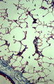 |
Peripheral alveolar area of the lung (human) | Stain: Azan. (1) Part of a pulmonary artery in a septum (2). (3) represents part of a bronchiolus respiratorius that continues into several alveolar ducts (4) and subsequently in alveolar sacs. Arrows (↓) indicate small foci of carbon deposits. | Respiratory bronchioli; Alveolar ducts; Alveolar sacs | Poja Histology Collection - Respiratory System Subset |
| 386 |
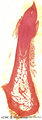 |
Permanent tooth - canine, human, adult; low magnification of labiolingual section | Stain: Hematoxylin and scarlet red. From top to bottom: Crown region with dentin but without enamel (decalcified specimens); Neck region at the attachment of the gingiva to dentin (left and right); Cementum is visible as a dark small rim from the neck region to the bottom of the tooth; Periodontal l... | oral cavity; alveolar process | Poja Histology Collection - Oral Cavity Subset |
| 387 |
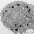 |
Peroxidase activity in neutrophilic granulocyte (peripheral blood, guinea pig) | Electron microscopy (peroxidase reaction with diaminobenzidin staining). The elongated nucleus (3) of this strongly ameboid phagocytic cell has several projections. These nuclear extensions are interconnected by thin heterochromatin strands. The cytoplasm exhibits moderate amounts of organelles, gly... | Poja Histology Collection - Blood & Bone Marrow Subset | |
| 388 |
 |
Peroxidase activity in neutrophilic myelocyte (postnatal liver, rat) | Electron microscopy (peroxidase reaction with diaminobenzidin staining). An early neutrophilic myelocyte with a large nucleus and well developed organelles, distinct Golgi areas (3) and granules. The Golgi areas, rough endoplasmic reticulum and nuclear membrane stain positively (species-dependent), ... | Poja Histology Collection - Blood & Bone Marrow Subset | |
| 389 |
 |
Peroxidase reaction in reticular cell and myelocytes (postnatal liver, rat) | Electron microscopy (peroxidase reaction with diaminobenzidin staining). The elongated reticular cell (1) shows peroxidase activity within the perinuclear space as well as the rough endoplasmic reticulum (species-dependent). It is surrounded by three eosinophilic myelocytes (2) with a positive react... | Poja Histology Collection - Blood & Bone Marrow Subset | |
| 390 |
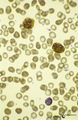 |
Peroxidase staining of granulocytes in peripheral blood smear (human) | Stain: peroxydase staining with DAB (diaminobenzidin). Blood smear showing two strongly positively stained granulocytes and one negative lymphocyte. The (myelo-) peroxidase activity is localized in the granules. | Poja Histology Collection - Blood & Bone Marrow Subset | |
| 391 |
 |
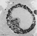
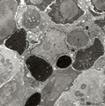
Phagocytosis in small splenic blood vessel (mouse) | Electron microscopy. Stain: Peroxidase reaction with diaminobenzidin staining. A diversity of red blood cells is black-stained due to the staining of hemoglobin by oxidized benzidin. Circulating lymphocytes (2) in the lumen (*) remain unstained. An oblong monocyte (1) developing into a macrophage h... | electron microscopy; phagocytosis | Poja Histology Collection - Lymphatic Tissues and Organs Subset |
| 392 |
 |
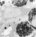
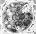
Phagocytosis in splenic red pulp (mouse) | Electron microscopy. Stain: Peroxidase reaction with diaminobenzidin staining. A diversity of red blood cells in the red pulp can be discerned due to the DAB staining of hemoglobin by oxidized benzidin (dark and light staining). The macrophage (1) shows peroxidase activity along the nuclear membran... | electron microscopy; phagocytosis | Poja Histology Collection - Lymphatic Tissues and Organs Subset |
| 393 |
 |
Phagocytosis of latex by neutrophil (peripheral blood, human) | Electron microscopy. The primary function of neutrophilic granulocytes is phagocytosis, ingestion of and destroying microbes or, as shown here, of latex particles. Several electron-light latex particles (1) (with a dense core) are internalized after incubation of free circulating granulocytes with a... | Poja Histology Collection - Blood & Bone Marrow Subset | |
| 394 |
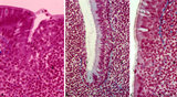 |
Pharyngeal tonsil ('gut-associated lymphatic tissue' or GALT) (human) | Stain: Azan. The combination demonstrates stages of infiltration (=diapedesis) of lymphocytes in the epithelium of the pharyngeal tonsil. The pharyngeal tonsil or adenoid is located in the nasopharyngeal roof. The agglomerations of lymphocytes (4) are covered by (1) pseudostratified ciliated epithel... | pharyngeal tonsil ; GALT; diapedesis; pseudostratified ciliated epithelium | Poja Histology Collection - Lymphatic Tissues and Organs Subset |
| 395 |
 |
Pharyngeal tonsil ('lymphoepithelial tissues', 'gut-associated lymphatic tissue' or GALT) (human) | Stain Azan. The solitary pharyngeal tonsil is localized in the pharyngeal fornix and belongs to the so-called Waldeyer's ring of pharyngeal lymphatic tissue. A: The roof of the nasopharynx is covered by a columnar epithelium with faintly light-stained goblet cells (1) and part of a fold shows cle... | pharyngeal tonsil; GALT; pseudostratified columnar epithelium | Poja Histology Collection - Lymphatic Tissues and Organs Subset |
| 396 |
 |
Plasma cell | Scheme electron microscopy. The mature plasma cell or plasmacyte (12-15 m) shows an eccentric nucleus (1) with a characteristic chunky distribution of heterochromatin along the inner nuclear membrane ('spoke-wheel' effect in light microscopy). Juxta-nuclearly an elaborate Golgi area (2) with secreti... | Poja Histology Collection - Blood & Bone Marrow Subset | |
| 397 |
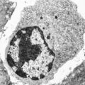 |
Plasma cell (liver, rat) | Electron microscopy. The mature plasma cell or plasmacyte shows an eccentric nucleus with a characteristic chunky distribution of heterochromatin along the inner nuclear membrane (spoke-wheel effect in light microscopy). Juxtanuclearly an elaborate Golgi area (2) (cytocentrum/centrosome). The cytopl... | Poja Histology Collection - Blood & Bone Marrow Subset | |
| 398 |
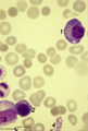 |
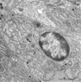
Plasma cell in bone marrow smear (human) | Stain: May-Grnwald-Giemsa (MGG). (1) A plasma cell with vacuoles (containing Ig), basophilic cytoplasm and a clear halo (Golgi apparatus) in a bone marrow film. (2) represents a young polychromatic erythroblast. (3) neutrophilic myelocyte, and (4) is a cluster of platelets. | Poja Histology Collection - Blood & Bone Marrow Subset | |
| 399 |
 |
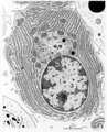
Plasma cell with Russell bodies (nose septum, rat) | Electron microscopy. This mature plasma cell exhibits in the cytoplasm dilated rough endoplasmic reticulum (1) filled with electron-grey material as well as electron-dense granules (2) of varying sizes (antibodies). These electron-dense granules are made up of accumulated (crystallin) immunoglobulin... | Poja Histology Collection - Blood & Bone Marrow Subset | |
| 400 |
 |
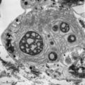
Plasma cells (spleen, mouse) | Electron microscopy. The mature plasma cell shows an eccentric nucleus with a characteristic chunky distribution of heterochromatin along the inner nuclear membrane ('spoke-wheel' effect in light microscopy). Juxta-nuclearly an elaborate Golgi area (2) with secretion vacuoles is localized (cytocentr... | Poja Histology Collection - Blood & Bone Marrow Subset |