The Health Education Assets Library (HEAL) is a collection of over 22,000 freely available digital materials for health sciences education. The collection is now housed at the University of Utah J. Willard Marriott Digital Library.
TO
Filters: Collection: "ehsl_heal"
| Title | Description | Subject | Collection | ||
|---|---|---|---|---|---|
| 376 |
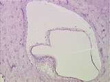 |
Ear | This image shows the bony labyrinth filled with perilymph, containing the membranous labyrinth filled with endolymph. Within the ampulla of each semicircular canal is the crista ampullaris, which is a specialization of the membranous labyrinth. UCLA Histology Collection. | Crista Ampullaris; Ear | UCLA Histology |
| 377 |
 |
Cells / Organelles - Developing Bone | Another area of the newborn rat knee, demonstrating a portion of developing bone, stained pinkish. The cells producing the bone are called osteoblasts. Identify a nucleus, nucleolus. The Golgi apparatus can be identified as the clear, central area. UCLA Histology Collection. | UCLA Histology | |
| 378 |
 |
Ingrown nail | 90% phenol is one of the agents advocated for destroying the nail matrix that grows at the base of the cul-de-sac beneath the proximal nail fold. | Knowledge Weavers Dermatology | |
| 379 |
 |
Uterus | In the ischaemic phase of the menstrual cycle the secretory endometrial glands begin to degenerate and there is intraendometrial hemorrhage. UCLA Histology Collection. | ischaemic endometrium | UCLA Histology |
| 380 |
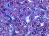 |
Cells / Organelles - Liver | In this slide, the liver cell cytoplasm appears red-purple. Identify nuclei and collagenous extracellular matrix (ECM), stained blue. UCLA Histology Collection. | UCLA Histology | |
| 381 |
 |
Uterine rupture | Uterine rupture | Knowledge Weavers Human Reproduction | |
| 382 |
 |
Lung carcinoma | Lung carcinoma | Knowledge Weavers Pathology | |
| 383 |
 |
Eye - Ciliary Epithelium | Some features of the double-layered ciliary epithelium can be observed in the plars plana region. The innermost layer (closest to the posterior chamber) is non-pigmented and the outermost layer is pigmented. The ciliary epithelium forms aqueous humor and secretes it into the posterior chamber. Aqueo... | canal of Schlemm; ciliary epithelium | UCLA Histology |
| 384 |
 |
Eye - Ciliary Body | The ciliary body is a ring-shaped structure that extends around the eyeball, posterior to the corneal - scleral junction (limbus). It consists of a double-layered epithelium, smooth muscle and stroma. Canal of schlemm is a landmark for the limbal region. The epithelium covers a flat portion of the c... | canal of schlemm | UCLA Histology |
| 385 |
 |
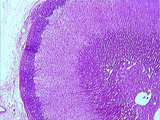
spleen | Reticular fibers are numerous in this image of red pulp. Reticular fibers encircle sinusoids like metal rings around a barrel. Depending on the angle of section, reticular fibers may appear as black dots, rings, or lines along the outer edge of the sinusoids. Identify sinusoids cut longitudinally an... | red pulp; reticular fibers; spleen | UCLA Histology |
| 386 |
 |
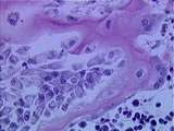
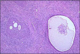
Adrenal | The classical organization of the adrenal gland with a thick capsule and the cortex composed of three layers: the outer zona glomerulosa, the middle zona fasciculata and the inner zona reticularis. Finally, in the center of the gland is the adrenal medulla. UCLA Histology Collection. | Adrenal | UCLA Histology |
| 387 |
 |
Skin - Hair Follicle | Associated with hair follicles are sebaceous glands and arrector pili muscles. The sebum produced by the glands is a holocrine secretion that is emptied onto the hair shaft. The arrector pili muscles pull the hair shaft into a more upright position. Identify adipose and hair. UCLA Histology Collecti... | UCLA Histology | |
| 388 |
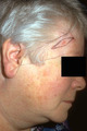 |
Basal cell carcinoma: excision removal | This person had a basal cell carcinoma, and the epidermis and dermis were excised. The danger area where the temporal branch of the facial nerve may lie and be quite close to the undersurface of the skin is shown by the straight lines drawn from ear to forehead. The surgeon should be careful to unde... | Surgical Methods | Knowledge Weavers Dermatology |
| 389 |
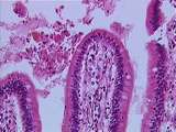 |
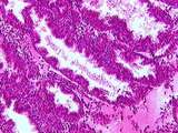
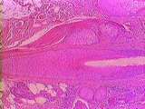
Gastrointestinal Tract | Low power view of a plica circularis of the jejunum; this is a fold of mucosa and submucosa. Many poorly preserved villi, evaginations of lamina propria and epithelium, can be seen protruding from the plica circularis. Invaginations of the epithelium into the lamina propria form intestinal glands ca... | gastrointestinal tract; plica circularis; small intestine | UCLA Histology |
| 390 |
 |
Brenner tumor | Solid and partially cystic epithelial nests are surrounded by a stroma composed of bundles of tightly-packed spindle shaped cells. The epithelial cells are polygonal and of the squamoid type, with pale, eosinophilic cytoplasm and oval nuclei having distinct nuclei and longitudinal grooving, a coffee... | Knowledge Weavers Human Reproduction | |
| 391 |
 |
Goodman | This is a picture of Dr. Goodman (of Goodman & Gillman fame). I once asked him how much you should know about a medication before using it, and he said,A lot! And you can quote me on that! | Knowledge Weavers Dermatology | |
| 392 |
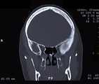 |
Fibrous Dysplasia | This image depicts Fibrous Dysplasia. Fibrous Dysplasia can be found in a radiograph and diagnosis is relatively easy if common symptoms are present. | McCune-Albright Syndrome; Lichtenstein Jaffe Disease; Caf-au-lait Spots; Leontiasis Ossium | HEAL Reviewed Collection |
| 393 |
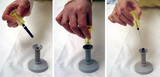 |
Minimize risks: procedure after venipuncture | A series of three images showing how to reduce the possibility of needle stick injuries after venipuncture by using an adapter. | Venipuncture | HEAL Reviewed Collection |
| 394 |
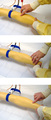 |
Search and find vein | Three examples of how to find and feel a vein and a good puncture spot. | Venipuncture; Vein Location | HEAL Reviewed Collection |
| 395 |
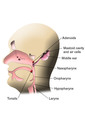 |
Pharynx (Labeled) | Pharyngeal structures. | Mastoid Cavity; Mastoid Air Cells | Royal College of Surgeons in Ireland Illustrations |
| 396 |
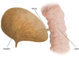 |
Bladder Fistula (Labeled) | Bladder Fistula. Colon. | Royal College of Surgeons in Ireland Illustrations | |
| 397 |
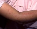 |
Rash of Systemic Onset Juvenile Rheumatoid Arthritis | Maculo-papular rash in a child with systemic onset rheumatoid arthritis (Still's Disease). The rash occurred during a flare-up. | Still's Disease; Systemic Onset; Maculo-papular Rash | HEAL Reviewed Collection |
| 398 |
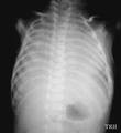 |
Anterior-posterior chest X-ray of meconium-stained severely asphyxiated infant | Anterior-posterior X-ray of meconium-stained severely asphyxiated infant with Type II Meconium Aspiration Syndrome (MAS) and shock lung (ARDS). | Anterior-Posterior Chest Diameter; Type II MAS; Shock Lung | Harris Pediatric Image Collection |
| 399 |
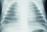 |
Anterior-posterior X-ray taken shortly after birth of infant with Type II MAS | Anterior-posterior chest X-ray taken shortly after birth of baby with Type II Meconium Aspiration Syndrome (MAS). | Anterior-Posterior Chest Diameter; Air Trapping; Type II MAS | Harris Pediatric Image Collection |
| 400 |
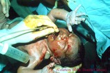 |
Further suctioning of MAS infant before first breath taken | Further suctioning of Meconium Aspiration Syndrome (MAS) infant before baby takes his/her first breath. | Mouth Suctioning | Harris Pediatric Image Collection |