The Health Education Assets Library (HEAL) is a collection of over 22,000 freely available digital materials for health sciences education. The collection is now housed at the University of Utah J. Willard Marriott Digital Library.
TO
| Title | Description | Subject | Collection | ||
|---|---|---|---|---|---|
| 376 |
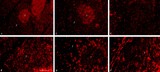 |
Immunohistochemistry with cellular markers in thymus (rat) | Stain: Alexa-594 red immunofluorescence. (1) medulla. (2) cortex. (A-survey, B-cortex): ER13 antibody stains for MHC-class II antigens on reticular cell types in medulla and cortex. (C): ED1 monoclonal antibody stains a single chain glycoprotein of 110 kDa on the lysosomal membrane of myeloid cel... | ER13 antibody; ED1 monoclonal antibody; ED2 monoclonal antibody; ER2 monoclonal antibody | Poja Histology Collection - Lymphatic Tissues and Organs Subset |
| 377 |
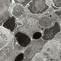 |
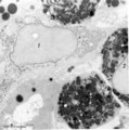
Phagocytosis in splenic red pulp (mouse) | Electron microscopy. Stain: Peroxidase reaction with diaminobenzidin staining. A diversity of red blood cells in the red pulp can be discerned due to the DAB staining of hemoglobin by oxidized benzidin (dark and light staining). The macrophage (1) shows peroxidase activity along the nuclear membran... | electron microscopy; phagocytosis | Poja Histology Collection - Lymphatic Tissues and Organs Subset |
| 378 |
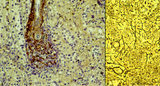 |
Red pulp of spleen with venous sinusoids (monkey, human) | Stain: A: Silver stain (Movat) (monkey); B: Silver stain (Gomori) (human). (A): The darkly stained fibres are conspicuous in the PALS (1) area arranged in parallel rows. The blood vessels continue in the surrounding splenic sinusoids (4). The wall of the sinusoid is built as a grid, the space is su... | sinusoid; reticular fibres | Poja Histology Collection - Lymphatic Tissues and Organs Subset |
| 379 |
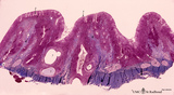 |
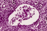
Survey of pharyngeal tonsil ('lymphoepithelial tissues', 'gut-associated lymphatic tissue' or GALT) (human) | Stain Azan. The solitary pharyngeal tonsil is localized in the pharyngeal fornix and belongs to the so-called Waldeyer's ring of pharyngeal lymphatic tissue. A survey shows the folded columnar epithelium (1) with faintly light-stained goblet cells with clefts (2) in between. The lamina propria co... | pharyngeal tonsil; GALT | Poja Histology Collection - Lymphatic Tissues and Organs Subset |
| 380 |
 |
Cystic corpuscle in thymus (human, adult) | Stain: Azan. With age the remaining Hassall's corpuscles become more prominent and might represent large multiform structures of over hundreds of microns. The central cells (*) are keratinized. They might be swollen, calcify and become necrotic eventually, or undergo lysis, leaving a large cystic st... | thymic corpuscle (Hassalls); epithelioreticular cell (ERC); lymphoid tissue | Poja Histology Collection - Lymphatic Tissues and Organs Subset |
| 381 |
 |
Peroxidase reaction in reticular cell and myelocytes (postnatal liver, rat) | Electron microscopy (peroxidase reaction with diaminobenzidin staining). The elongated reticular cell (1) shows peroxidase activity within the perinuclear space as well as the rough endoplasmic reticulum (species-dependent). It is surrounded by three eosinophilic myelocytes (2) with a positive react... | Poja Histology Collection - Blood & Bone Marrow Subset | |
| 382 |
 |
Plasma cell (liver, rat) | Electron microscopy. The mature plasma cell or plasmacyte shows an eccentric nucleus with a characteristic chunky distribution of heterochromatin along the inner nuclear membrane (spoke-wheel effect in light microscopy). Juxtanuclearly an elaborate Golgi area (2) (cytocentrum/centrosome). The cytopl... | Poja Histology Collection - Blood & Bone Marrow Subset | |
| 383 |
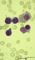 |
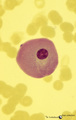
Three basophilic erythroblasts in bone marrow smear (human) | Stain: May-Grnwald-Giemsa (MGG). Three basophilic erythroblasts (1) with intense blue stained cytoplasm, and some so called ears or cytoplasmic projections (arrows). Chromatin strands are thicker than in the proerythroblast. Generally no nucleoli are seen. (2) Damaged or smudged eosinophilic myelocy... | Poja Histology Collection - Blood & Bone Marrow Subset | |
| 384 |
 |
Polychromatic erythroblasts and neutrophilic granulocytes in bone marrow smear (human) | Stain: May-Grnwald-Giemsa (MGG). Three polychromatic erythroblasts or normoblasts (1) and two neutrophils (2). The erythroblasts show light basophilic cytoplasm and well condensed nuclear chromatin. The lower neutrophil (more mature) has a more segmented nucleus than the upper one. | Poja Histology Collection - Blood & Bone Marrow Subset | |
| 385 |
 |
Erythron in bone marrow smear (human) | Stain: May-Grnwald-Giemsa (MGG). The erythron or erythroblastic island consists of a large reticulum cell (1) surrounded by erythroblastic cell types (2) at varoius stages of differentiation. The nucleus of the reticulum cell (histiocyte) usually contains a noticeable nucleolus. At distance, some my... | Poja Histology Collection - Blood & Bone Marrow Subset | |
| 386 |
 |
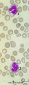
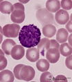
Mast cell in bone marrow smear (human) | Stain: May-Grnwald-Giemsa (MGG). The mast cell (1) is characterized by the presence of coarse purple-black granules and a nucleus with oval, central nucleus. The granules contain pharmacologically active mediators such as heparin, histamine, neutrophil- and eosinophil-chemotactic factors, vasoactive... | Poja Histology Collection - Blood & Bone Marrow Subset | |
| 387 |
 |
Degenerating granulocytes in peripheral blood smear (human) | Stain: May-Grnwald-Giemsa (MGG).Two neutrophils with degenerated nuclei. Note the pyknotic pattern (like mature erythroblast) of the separated and rounded nuclear lobes and the granules in the cytoplasm. Normally only a small number of granulocytes die in the blood. | Poja Histology Collection - Blood & Bone Marrow Subset | |
| 388 |
 |
Giant plasma cell in peripheral blood smear (human) | Stain: May-Grnwald-Giemsa (MGG). A giant plasma cell with diffusely spread immunoglobulins in the cytoplasm. The Golgi area remains unstained (white area). | Poja Histology Collection - Blood & Bone Marrow Subset | |
| 389 |
 |
Vacuolar degeneration of neutrophils in peripheral blood smear (human) | Stain: May-Grnwald-Giemsa (MGG). Toxic degeneration of neutrophil resulting in vacuolization of cytoplasm (1); cell with beginning vacuolization (2). | Poja Histology Collection - Blood & Bone Marrow Subset | |
| 390 |
 |
Activated lymphocyte (liver inflammation, human) | Electron microscopy. A cytotoxic T cell (2) is localized within the sinusoid (3) in the liver (8), surrounded by erythrocytes (1) and debris. The lymphocyte with an irregular indented nucleus shows juxta-nuclearly partly swollen Golgi areas (*) and aggregation of mitochondria (4) in the cytoplasm. F... | Poja Histology Collection - Blood & Bone Marrow Subset | |
| 391 |
 |
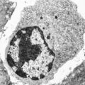
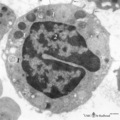
Monocyte (peripheral blood, human) | Electron microscopy. In this picture the large nucleus (1) of this cell (diameter of 12-20 μm) is twice sectioned. Golgi areas (2) and centriole, profiles of rough endoplasmic reticulum (3) and many free ribosomes are present. There are many mitochondria (4) as well as scattered homogeneous electro... | Poja Histology Collection - Blood & Bone Marrow Subset | |
| 392 |
 |
Erythroblasts (bone marrow, rabbit) | Electron microscopy. (1) shows basophilic erythroblasts or early normoblasts with clumped heterochromatin in the nucleus and numerous polysomes. (1m) points to a mitotic figure of a basophilic erythroblast. A late polychromatic erythroblast or intermediate normoblast (2) marks a maturing stage in wh... | Poja Histology Collection - Blood & Bone Marrow Subset | |
| 393 |
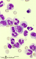 |
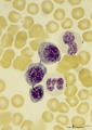
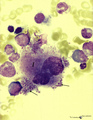
Myelopoiesis in bone marrow smear (human) | Stain: May-Grnwald-Giemsa (MGG). The smear shows a group of myeloid cells in different stages of maturation, in which series the alterations in the nucleus can be noticed clearly going from (1) to (5), i.e. the nucleus gets more and more condensed, indented and subsequently segmented in lobes. (1) p... | Poja Histology Collection - Blood & Bone Marrow Subset | |
| 394 |
 |
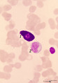
Basophilic granulocyte in peripheral blood smear (human) | Stain: May-Grnwald-Giemsa (MGG). The basophilic granulocyte has large, coarse dark purple-stained granules that contain mediators such as eosinophilic chemotactic factors, heparin, histamine, myeloperoxidase. The nuclear lobes are irregular, badly visible and covered by the granules. | Poja Histology Collection - Blood & Bone Marrow Subset | |
| 395 |
 |
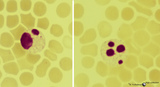
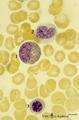
Band and hyper segmented neutrophils in peripheral blood smear (human) | Stain: May-Grnwald-Giemsa (MGG). (A) shows a young band neutrophil with a non-segmented nucleus. (B) shows mature neutrophils with more than 6 nuclear lobes (hyper segmented). The smear also shows anisocytosis with both microcytes and macrocytes. | Poja Histology Collection - Blood & Bone Marrow Subset | |
| 396 |
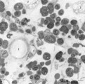 |
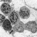
Survey of bone marrow (bone marrow, rabbit) | Electron microscopy. Within the loose reticular tissue a great variety of different stages of young erythrocytes (3, 4) as well as of young granulocytes (5) is present. (2) indicates reticular cells and (2b) phagocytizing reticular cells. (1) fat cells (adipocytes); (6) myeloid cell; (7) capillary. | Poja Histology Collection - Blood & Bone Marrow Subset | |
| 397 |
 |
Eosinophilic (meta)myelocyte in bone marrow smear (human) | Stain: May-Grnwald-Giemsa (MGG). The granules of the eosinophilic (meta)myelocyte (1) are large and brown-blue stained in contrast to the hardly visible, dust-like granules in the neutrophilic band form (2). (3) orthochromatic erythroblast. | Poja Histology Collection - Blood & Bone Marrow Subset | |
| 398 |
 |
Granular megakaryocyte producing platelets in bone marrow smear (human) | Stain: May-Grnwald-Giemsa (MGG). The centrally located megakaryocyte (1) has a huge nucleus (polyploidy) and an extended cytoplasm from which platelets are released at the periphery (arrows). | Poja Histology Collection - Blood & Bone Marrow Subset | |
| 399 |
 |
Mast cell (lung, dog) | Electron microscopy. This mucosal mast cell of the lung is localized in the vicinity of a blood vessel. Notice the smooth muscle cells (1) of a small arteriole and collagen fibers (2). Most obvious is the presence of granules varying in shape and size. These membrane-bound vesicles (so-called compou... | Poja Histology Collection - Blood & Bone Marrow Subset | |
| 400 |
 |
Basophilic granulocyte (peripheral blood, human) | Electron microscopy. The bilobed nucleus (2) is surrounded by moderate amount of organelles. The cell exhibits few short filopodia (arrows). The large coarse basophilic granules (1) (specific granules) vary in density and contain vasoactive mediators such as heparin, histamine, sulphated proteoglyca... | Poja Histology Collection - Blood & Bone Marrow Subset |