The Health Education Assets Library (HEAL) is a collection of over 22,000 freely available digital materials for health sciences education. The collection is now housed at the University of Utah J. Willard Marriott Digital Library.
TO
Filters: Collection: ehsl_heal
| Title | Description | Subject | Collection | ||
|---|---|---|---|---|---|
| 351 |
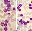 |
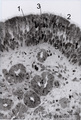
Normal pleiomorph and leukemic monomorph bone marrow smear (human) | Stain: May-Grnwald-Giemsa (MGG). In normal bone marrow smear (A) a pleimorph or heterogeneous collection of different cell types (A1-7) can be identified, while in the bone marrow smear of a leukemic patient a single type dominates (in this case lymphoid cells, B-1). | Poja Histology Collection - Blood & Bone Marrow Subset | |
| 352 |
 |
Normal proerythroblasts in bone marrow smear (human) | Stain: May-Grnwald-Giemsa (MGG). The three mounted proerythroblasts are easily recognized by their so called ears i.e., the blue cytoplasmic projections or extensions (arrows). The basophilic cytoplasm as well as the nucleoli point to the blast character of the cells. | Poja Histology Collection - Blood & Bone Marrow Subset | |
| 353 |
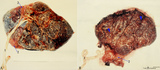 |
Normal term placenta (human) | (A) Left (fetal surface): 30-90 cm long umbilical cord (1), remnant of ruptured transparent amnion (2→) , near the eccentric attachment of the umbilical cord to the chorion plate ramification of the umbilical vessels (3) (arteries are smaller than the veins). (B) Right (maternal surface). The su... | placenta; amnion; chorion plate; cotyledon | Poja Histology Collection - Placenta |
| 354 |
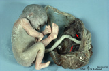 |
Normal term placenta with umbilical cord attached to the fetus (human) | The macroscopy shows the expelled placenta as a discoidal mass with a circular outline about 15-20 cm in the ruptured amnionitic and chorionic sacs. Amnion and smooth chorion (chorion leave) are fused and continuous with the margins of the placenta. The fetal surface (inner surface) is covered by... | placenta; amnion; chorion; umbilical cord; fetus | Poja Histology Collection - Placenta |
| 355 |
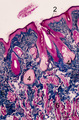 |
Nostril area of nasal vestibulum - cutaneous region - human | Stain: Azan. Slightly keratinized squamous epithelium (1) with thin red cornified layer; (2) hair follicles; (3) vibrissae and (4) associated sebaceous glands. At the bottom skeletal muscle fibers; (5) transverse part of the musculous nasalis; (6) connective tissue. | Poja Histology Collection - Respiratory System Subset | |
| 356 |
 |
Nucleated erythrocytes in peripheral blood smear (bird) | Stain: May-Grnwald-Giemsa (MGG). In contrast to human red blood cells mature erythrocytes of birds contain nuclei (1). (2) Indicates an eosinophilic granulocyte. Inset demonstrates chromatin pattern of the nuclei. | Poja Histology Collection - Blood & Bone Marrow Subset | |
| 357 |
 |
Nucleus expulsion in polychromatic-orthochromatic erythroblasts in bone marrow smear (human) | Stain: May-Grnwald-Giemsa (MGG). The composed pictures show three sequential stages in the final maturation stage of red blood cells. (A) polychromatic erythroblast with a condensed nucleus and an almost acidophilic cytoplasm. (B) the nucleus in the orthochromatic erythroblast is completely condense... | Poja Histology Collection - Blood & Bone Marrow Subset | |
| 358 |
 |
Odontoblast in tooth development - mammalian embryo | Scheme electronmicroscopy. A columnar and irregular shaped cell (mesenchymal origin) with a taperwise odontoblastic process within the predentin. The bottom side is adjacent to the future pulp cells. The cell body contains many organelles and especially a well-developed Golgi area with prosecretoy g... | oral cavity | Poja Histology Collection - Oral Cavity Subset |
| 359 |
 |
Olfactory epithelium in the nasal cavity (mammals) | Scheme electron microscopy. Four olfactory bulbs (1, vesicles) with radially sprouting cilia are present at the surface of the epithelium. Their slender cell bodies (2, bipolar neurons) are flanked by sustentacular (supporting) broad cells (3) whose apices are filled with well developed organelles, ... | Pseudostratified epithelium; Olfactory epithelium; Basal cells; Olfactory vesicle; Bipolar neuron | Poja Histology Collection - Respiratory System Subset |
| 360 |
 |
Olfactory epithelium in the nasal cavity (mammals) | Three-dimensional scheme electron microscopy. Six olfactory cells (1) surround one massive central supporting cell (2). A third, basal cell (3) is located at the basal plate aposing to the supporting cell. These olfactory cells are tall and slender bipolar nerve cells with chemoreceptive functions. ... | Pseudostratified epithelium; Olfactory epithelium; Basal cells; Olfactory vesicle; Bipolar neuron | Poja Histology Collection - Respiratory System Subset |
| 361 |
 |
Olfactory epithelium in the nose (human) | Electron microscopy. At the top (lumen) cross-sections of olfactory bulbs (1) (vesicles) with cilia/basal bodies. Sustentacular (supporting) cells (2) exhibit numerous microvilli in the lumen, their cytoplasms contain many dispersed mitochondria and in the lower half aggregations of endoplasmic reti... | Olfactory epithelium; Olfactory vesicle | Poja Histology Collection - Respiratory System Subset |
| 362 |
 |
Olfactory gland (Bowman) in olfactory region of concha (human) | Electron microscopy. Beneath the olfactory epithelium tubuloalveolar glands of Bowman (1) are found containing cells that produce dense secretion granules arranged around a large lumen (*) with greyish secretion product. These cells produce a serous fluid in which odoriferous substances are dissolve... | Olfactory epithelium; Bowman glands; Olfactory glands; Secretion granules | Poja Histology Collection - Respiratory System Subset |
| 363 |
 |
Olfactory mucosa in the nose (dog) | Stain: Iron Hematoxylin and eosin (Heidenhain). The pseudostratified epithelium of the olfactory mucosa contains sensory cells, support cells and basal cells. The upper darker stained nuclei (1) belong to the supporting (sustentacular) cells showing a dark apical line (↑) at the top. Below lighte... | Bowman glands; Olfactory glands; Olfactory mucosa; Pseudostratified epithelium | Poja Histology Collection - Respiratory System Subset |
| 364 |
 |
Olfactory mucosa in the nose (human) | Stain: Toluidine blue, one-micron Epon plastic section. Pseudostratified epithelium with light stained nuclei (1) (note distinct nucleoli) of olfactory cells. At the surface faintly stained cilia/microvilli (2). Nuclei of sustentacular (supporting) cells (3) are stained darker and predominantly in t... | Bowman glands; Olfactory glands; Olfactory mucosa; Pseudostratified epithelium | Poja Histology Collection - Respiratory System Subset |
| 365 |
 |
Olfactory vesicle (bulb) in the nose (gerbil) | DUPLICATE RECORD - FOR DELETION Electron microscopy. A distinct olfactory bulb (1, vesicle) with horizontally extended cross-sectioned cilia (*) and basal bodies (↑) is surrounded by numerous microvilli and free olfactory cilia. Close to the first bulb a second one is about to protrude above th... | Olfactory epithelium; Olfactory vesicle | Poja Histology Collection - Respiratory System Subset |
| 366 |
|
Olfactory vesicle (bulb) in the nose (gerbil) | Electron microscopy. A distinct olfactory bulb (1, vesicle) with horizontally extended cross-sectioned cilia (*) and basal bodies (↑) is surrounded by numerous microvilli and free olfactory cilia. Close to the first bulb a second one is about to protrude above the surface of the supporting cells b... | Olfactory epithelium; Olfactory vesicle | Poja Histology Collection - Respiratory System Subset |
| 367 |
 |
Orthochromatic erythroblast (bone marrow, rabbit) | Electron microscopy. The orthochromatic erythroblast (1) or late normoblast shows the early stage of extrusion of the nucleus though the latter appears not yet fully pyknotic. The cytoplasm contains few slender mitochondria (2) as well as scattered clumped polysomes. (3) Part of a reticulocyte. | Poja Histology Collection - Blood & Bone Marrow Subset | |
| 368 |
 |
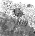
Orthochromatic erythroblast (bone marrow, rabbit) | Electron microscopy. The orthochromatic erythroblast or late normoblast is surrounded by young granulocytes (3) and part of a megakaryocyte (2). The cell shows the early stage of extrusion of the nucleus (1) that is already slightly pyknotic. The cytoplasm contains few small mitochondria (arrows) an... | Poja Histology Collection - Blood & Bone Marrow Subset | |
| 369 |
 |
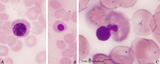
Orthochromatic erythroblast and lymphocyte in bone marrow smear (human) | Stain: May-Grnwald-Giemsa (MGG). The cytoplasm of this orthochromatic erythroblast (2) shows a faint polychromatic tint. The chromatin is arranged in clumps and stains deeply. (2) activated lymphocyte. | Poja Histology Collection - Blood & Bone Marrow Subset | |
| 370 |
 |
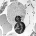
Orthochromatic erythroblast in bone marrow smear (human) | Stain: May-Grnwald-Giemsa (MGG). The orthochromatic erythroblast (1) has a dark condensed (pyknotic) nucleus located at one site of the cell, ready for being expulsed from the cell. (2) Erythrocyte. (3) Represents a platelet or thrombocyte. | Poja Histology Collection - Blood & Bone Marrow Subset | |
| 371 |
 |
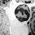
Orthochromatic erythroblasts in bone marrow smear (human) | Stain: May-Grnwald-Giemsa (MGG). Two orthochromatic erythroblasts (1) with condensed nuclear chromatin and a transition of cytoplasmic stain towards the color of normal erythrocytes. (2) a segmented neutrophilic granulocyte. | Poja Histology Collection - Blood & Bone Marrow Subset | |
| 372 |
 |
PAS-positively stained neutrophilic granulocytes in bone marrow smear (human) | Stain: Periodic acid-Schiff reaction (PAS). Mature neutrophils (1) have fine positive granules within negatively (un)stained cytoplasm, whereas eosinophils (2) and basophils show a positive cytoplasmic reaction contrasting with the negative granules. In lymphocytes (1) PAS-positive granules are rare... | Poja Histology Collection - Blood & Bone Marrow Subset | |
| 373 |
 |
Palatine tonsil ('lymphoepithelial tissues', 'gut-associated lymphatic tissue' or GALT) (human) | Stain: Azan. A low magnification in A shows the thick blue connective septum (1) with a secondary lymphatic nodule (2). (3) is a cross-section of a crypt filled with detached squamous epithelium (3a) mixed up with keratinized material (red) and a huge amount of lymphocytes (3b). A higher magnificati... | follicle; germinal center; mantle layer; stratified squamous epithelium | Poja Histology Collection - Lymphatic Tissues and Organs Subset |
| 374 |
 |
Papillae circumvallatae of the tongue (dorsal side, human) | Stain: Azan. Three broad papillae with taste buds facing the grooves in which the serous von Ebner glands drain. Striated skeletal muscles (musculus verticalis linguae and musculus longitudinalis superior) and lightly stained mucous glands (posterior lingual glands). | oral cavity; von Ebner; lingual muscles; lingual glands | Poja Histology Collection - Oral Cavity Subset |
| 375 |
 |
Papillae circumvallatae of the tongue (dorsal side, human) | Stain: Azan. A broad papilla with taste buds (lightly stained spots) facing the grooves in which the serous glands (von Ebner) drain. | oral cavity; von Ebner; circumvallate papillae | Poja Histology Collection - Oral Cavity Subset |