The Health Education Assets Library (HEAL) is a collection of over 22,000 freely available digital materials for health sciences education. The collection is now housed at the University of Utah J. Willard Marriott Digital Library.
Filters: Collection: ehsl_heal
| Title | Description | Subject | Collection | ||
|---|---|---|---|---|---|
| 326 |
 |
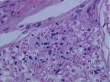
Peripheral Nervous System | A rather low power magnification of autonomic ganglion cells. Note the eccentric nuclei and the irregularly shaped cell bodies. These cells are efferent (motor) cells of the autonomic nervous system. Note the relatively sparse satellite cells in this image, and the open space surrounding the ganglio... | autonomic ganglion; Peripheral Nervous System | UCLA Histology |
| 327 |
 |
Peripheral Nervous System | This longitudinal section of myelinated peripheral nerve is stained with Kornhauser's Quad and should be compared to slide N9-40-1. Here we can visualize the dark-stained axon, located centrally within the light-stained myelin sheath. Note that the axon is continuous across the node of Ranvier. UCLA... | Kornhauser's Quad; peripheral nerve; Peripheral Nervous System | UCLA Histology |
| 328 |
 |
Peripheral Nervous System | The trigeminal ganglion, like a dorsal root ganglion, contains the cell bodies of sensory neurons. Identify these ganglion cells. Note the bundles or fascicles of nerves, and how the connective tissue capsule (epineurium) surrounds the ganglion. Identify the vein. UCLA Histology Collection. | epineurium; ganglion; Peripheral Nervous System | UCLA Histology |
| 329 |
 |
Peripheral Nervous System | Longitunial section of myelinated peripheral nerve. Axons are stained, myelin is not. Also see the perineurium. UCLA Histology Collection. | Peripheral Nervous System | UCLA Histology |
| 330 |
 |
Peripheral Nervous System | This cross section of a peripheral nerve demonstrates the appearance of myelinated axons and a bundle of unmyelinated axons. Unmyelinated axons in the PNS are embedded into the Schwann cells; the Schwann cells and associated axons constitute a REMAK bundle. The nerve is surrounded by epineurium. Wit... | Peripheral Nervous System; PNS; REMAK bundle | UCLA Histology |
| 331 |
|
Peripheral Nervous System | This cross section of a peripheral nerve demonstrates the appearance of myelinated axons and a bundle of unmyelinated axons. Unmyelinated axons in the PNS are embedded into the Schwann cells; the Schwann cells and associated axons constitute a REMAK bundle. The nerve is surrounded by epineurium. Wit... | Peripheral Nervous System; PNS; REMAK bundle | UCLA Histology |
| 332 |
 |
Peripheral Nervous System / Central Nervous System | The relationship between the spinal cord, the spinal roots, and the meninges is illustrated here. The surface of the spinal cord is covered by pia mater. Between the pia mater and web-like arachnoid mater is the subarachnoid space which contains CSF. The thick, collagenous dura mater is continuous w... | arachnoid mater; Central Nervous System; dura mater; meninges; Peripheral Nervous System; pia mater; spinal cord; spinal roots | UCLA Histology |
| 333 |
 |
Pineal Gland | Identify the corpora arenacea (brain sand) which are diagnostic of the pineal. Corpora arenacea are radio-opaque, which allows for identification of the pineal gland in X-rays and CT scans of the skull. UCLA Histology Collection. | corpora arenacea (brain sand); Pineal gland | UCLA Histology |
| 334 |
 |
Pineal Gland | This is a low magnification image of the pineal gland. You are not responsible for recognizing the cells of the pineal, but you are responsible for recognizing corpora arenacea or Brain Sand which are shown at higher magnification in image 167-25-1. Note: many people ingest melatonin, the product of... | Pineal gland | UCLA Histology |
| 335 |
|
Pineal Gland | This is a low magnification image of the pineal gland. You are not responsible for recognizing the cells of the pineal, but you are responsible for recognizing corpora arenacea or Brain Sand which are shown at higher magnification in image 167-25-1. Note: many people ingest melatonin, the product of... | Pineal gland | UCLA Histology |
| 336 |
 |
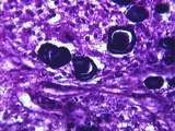
Pituitary | Because of its physiological importance, several images of the pars distalis are included. In this one, recognize bluish basophils, abundant reddish acidophils, a probable chromophobe (with no cytoplasmic granules), and red blood cells. UCLA Histology Collection. | pars distalis; Pituitary | UCLA Histology |
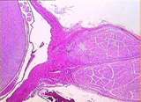
| 337 |
 |
Pituitary | Relatively low power view of the pars distalis abutting against the pars nervosa. There are some hypophyseal cysts shown. UCLA Histology Collection. | Pituitary | UCLA Histology |
| 338 |
 |
Pituitary | The pituitary stalk consists of the pars tuberalis (with RBC-filled portal vessels) and the neural stalk (the axons of the hypothalamo - hypophyseal tract). Both the pars tuberalis and the pars nervosa transport hormonal factors; can you name these hormones? UCLA Histology Collection. | Pituitary; pituitary stalk | UCLA Histology |
| 339 |
 |
Pituitary | This image shows an unusually large hypophyseal cyst surrounded by a shrinkage artifact and invading basophils from the former region of the pars intermedia which are now within the pars nervosa. A higher magnification of this region is found in image 166-40-2. UCLA Histology Collection. | Pituitary | UCLA Histology |
| 340 |
 |
Pituitary | In this image of the pars distalis, which is stained with a special PAS-Trichrome preparation, the basophils and acidophils appear less intermingled. What hormones do these two cell types secrete into the sinusoids? UCLA Histology Collection. | PAS-Trichrome; Pituitary | UCLA Histology |
| 341 |
 |
Pituitary | This is a coronal slice reconstructed by computer technology to demonstrate the median eminence of the hypothalamus which lies inferior to the third ventricle. Certain neurons of the median eminence terminate adjacent to capillary loops which drain into portal vessels. These portal vessels of the pa... | Pituitary | UCLA Histology |
| 342 |
 |
Pituitary | This sagittal slice shows the neural stalk as it turns posteriorly to form the pars nervosa. The neural stalk is made up of axons from the supraoptic and paraventricular nuclei of the hypothalamus. One can clearly recognize the difference between the pars distalis and the neural tissue. Also, an hyp... | Pituitary; pars nervosa | UCLA Histology |
| 343 |
 |
Pituitary | This sagittal slice shows the components of the pituitary stalk: the pars tuberalis and the neural stalk. The hypophyseal portal vessels of the pars tuberalis can be seen here in longitudinal section. The neural stalk is composed of axons which terminate in the pars nervosa (posterior pituitary). UC... | Pituitary; pituitary stalk | UCLA Histology |
| 344 |
 |
Pituitary | This oil immersion image of the pars distalis shows acidophils, basophils, and probable chromophobes. The chromophobes contain few or no stained cytoplasmic granules. UCLA Histology Collection. | Pituitary | UCLA Histology |
| 345 |
 |
Pituitary | Invading basophils from the pars intermedia located within the pars nervosa. Note that the special polychrome stain produces multicolored red blood cells, as seen in this branching blood vessel. Also identify the small, pale nuclei of pituicytes. UCLA Histology Collection. | basophilic invasion; basophils; pars intermedia; Pituitary | UCLA Histology |
| 346 |
 |
Pituitary | This image readily differentiates the endocrine tissue of the pars distalis and the nervous tissue of the neural stalk. The pars distalis receives a rich supply of blood via sinusoids. UCLA Histology Collection. | pars distalis; Pituitary | UCLA Histology |
| 347 |
 |
Pituitary | Pars nervosa consisting of nerve fibers, pituicytes and blood vessels. this special polychrome stain produces multi-colored red blood cells. Contrast with pars distalis. UCLA Histology Collection. | Hypophysis; Pituitary | UCLA Histology |
| 348 |
 |
Pituitary | A high magnification image of the pars distalis shows the rich blood supply via sinusoids and the staining difference between the cytoplasmic granules of acidophils and basophils. What is the chemical basis of the staining characteristics of these chromophil cells? For higher power view see 166-100-... | pars distalis; Pituitary | UCLA Histology |
| 349 |
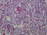 |
Placenta | This image presents the basic organization of the placenta. Embedded within the maternal blood space are numerous fetal villi, some of them clearly tertiary villi , all lined by the syncytiotrophoblast . The only things missing in this image are maternal decidual cells and fetal/maternal fibrinoid. ... | Placenta | UCLA Histology |
| 350 |
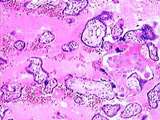 |
Placenta | This image presents all that you have to know about placental histology for this course. Fetal villi, some clearly tertiary villi , are completely surrounded by the maternal blood space. Lining everything in this image is the syncytiotrophoblast . The only other maternal component in this image is a... | Placenta | UCLA Histology |