The Health Education Assets Library (HEAL) is a collection of over 22,000 freely available digital materials for health sciences education. The collection is now housed at the University of Utah J. Willard Marriott Digital Library.
TO
Filters: Collection: "ehsl_heal"
| Title | Description | Subject | Collection | ||
|---|---|---|---|---|---|
| 326 |
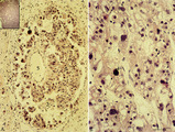 |
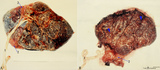
Choriocarcinoma (human) | Stain: Hematoxylin-eosin. (A) Inset: Survey tumor. Within the uterus (1) a choriocarcinoma forms solitary or multiple nodules composed of hemorrhagic necrotic areas (2) surrounded by neoplastic cells. It resembles an early implanted blastocyst with aggregations of mononuclear lighter-stained cyt... | choriocarcinoma; placenta; uterus; cytotrophoblast; GTD (gestational trophoblastic disease) | Poja Histology Collection - Placenta |
| 327 |
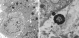 |
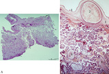
Electron microscopy of tertiary villus (human placenta, midpregnancy) | In the left photograph (A) is shown part of a tertiary villus with the organelle-rich cytoplasm of a syncytiotrophoblast cell (STC, 1). Below a single electron-light linked cytotrophoblast cell (CTC or Langhans cell, 2) covered by the STC. A higher magnification of another CTC (right photograph) sh... | placenta; tertiary villi; syncytiotrophoblast; electron microscopy; placental barrier | Poja Histology Collection - Placenta |
| 328 |
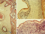 |
Chorioamnionitis (human) | Histologically chorioamnionitis describes the progression of the inflammatory process. Bacteria firstly colonized the chorioamniotic surface. In first two days polymorphonuclear granulocytes (PMN) migrate to the chorion (chorionitis) marginate and adhere to the bottom of the chorionic plate (stage 1... | chorioamnionitis; placenta; trophoblast | Poja Histology Collection - Placenta |
| 329 |
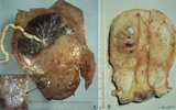 |
Partial hydatidiform mole and invasive mole (human) | (A) Macroscopy: The partial mole (1) occupies a large part of the placenta and is distinct from the normal chorionic plate where the umbilical cord (2) inserts eccentrically with branches of the umbilical vessels. The inset shows a circumscript area with swollen transparent grape-like vesicles (chor... | partial hydatidiform mole ; chorioadenoma | Poja Histology Collection - Placenta |
| 330 |
 |
Normal term placenta (human) | (A) Left (fetal surface): 30-90 cm long umbilical cord (1), remnant of ruptured transparent amnion (2→) , near the eccentric attachment of the umbilical cord to the chorion plate ramification of the umbilical vessels (3) (arteries are smaller than the veins). (B) Right (maternal surface). The su... | placenta; amnion; chorion plate; cotyledon | Poja Histology Collection - Placenta |
| 331 |
 |
Survey and detail of the chorionic plate and intervillous space (human placenta, full-term) | Stain: (A) Perjodic acid-Schiff reaction (PAS); (B) Hematoxylin-azophloxine. (A) At the top the chorionic plate (1) with cross-sections of umbilical vessels (2). At (3) the folded amnion covering the chorionic plate. Ramifications of thicker stem villi (6) demonstrate free-floating terminal villi ... | placenta; chorionic plate; amnion; terminal villi | Poja Histology Collection - Placenta |
| 332 |
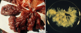 |
Fetal cotyledons (normal term human placenta) | After dissection of one placental lobe several cotyledons (2) are visible in (A). Each fetal cotyledon (2) consists of a main stem villus (1) and all its branches. (B) After trypsinization of a cotyledon the tree of arborisation of the stem villus and its branches becomes visible. (By courtesy... | placenta; cotyledon; villus; trypsinization | Poja Histology Collection - Placenta |
| 333 |
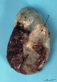 |
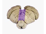
Macroscopy of fetus (human) | The fetus (1)is completely wrapped in a shiny transparent amnion (2) and closely associated with the brown-coloured placenta (3). (By courtesy of the Museum of Anatomy and Pathology, University Medical Center, St. Radboud University, Nijmegen, The Netherlands) | fetus; placenta; amnion | Poja Histology Collection - Placenta |
| 334 |
 |
Coordination Exam: Anatomy: Exam Tests (x2) (includes Spanish audio & captions) | The following tests of the neuro exam can be divided according to which system of the cerebellum is being examined: Vestibulocerebellum and spinocerebellum (midline): - Station - Walking - Tandem gait Cerebrocerebellum (appendicular): - Rapid alternating movements - Finger-to-nose - To... | Coordination Examination | NeuroLogic Exam: An Anatomical Approach |
| 335 |
 |
Coordination Exam: Normal Exam: Check Reflex (x2) (includes Spanish audio & captions) | Examiner pulls on actively flexed arm then suddenly releases. The patient should be able to check or stop the arm's movement when released. NeuroLogic Exam has been supported by a grant from the Slice of Life Development Fund at the University of Utah, the Department of Pediatrics and the Office of ... | Coordination Examination; Check Reflex | NeuroLogic Exam: An Anatomical Approach |
| 336 |
 |
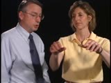
Coordination Exam: Normal Exam: Speech - Rapid Alternating Movements (x2) (includes Spanish audio & captions) | Having the patient say lah-pah-kah can test rapid alternating movements of the tongue, lips, and palate. NeuroLogic Exam has been supported by a grant from the Slice of Life Development Fund at the University of Utah, the Department of Pediatrics and the Office of Education at the University of Nebr... | Coordination Examination; Rapid Alternating Movements | NeuroLogic Exam: An Anatomical Approach |
| 337 |
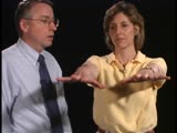 |
Coordination Exam: Normal Exam: Rebound (x2) (includes Spanish audio & captions) | Tap outstretched arms. Patient's arms should recoil to original position. NeuroLogic Exam has been supported by a grant from the Slice of Life Development Fund at the University of Utah, the Department of Pediatrics and the Office of Education at the University of Nebraska Medical Center. Viewing... | Coordination Examination; Rebound | NeuroLogic Exam: An Anatomical Approach |
| 338 |
|
Coordination Exam: Normal Exam: Rebound (includes Spanish audio & captions) | Tap outstretched arms. Patient's arms should recoil to original position. NeuroLogic Exam has been supported by a grant from the Slice of Life Development Fund at the University of Utah, the Department of Pediatrics and the Office of Education at the University of Nebraska Medical Center. Viewing... | Coordination Examination; Rebound | NeuroLogic Exam: An Anatomical Approach |
| 339 |
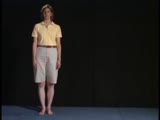 |
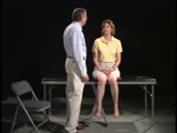
Gait Exam: Normal Exam: Station | The patient should be able to stand still with her feet less then shoulder width apart. NeuroLogic Exam has been supported by a grant from the Slice of Life Development Fund at the University of Utah, the Department of Pediatrics and the Office of Education at the University of Nebraska Medical Cent... | Gait Examination; Station | NeuroLogic Exam: An Anatomical Approach |
| 340 |
 |
Male Reproductive System | Anatomy of testicle. | Testicle; Glans Penis; Ductus Deferens | Royal College of Surgeons in Ireland Illustrations |
| 341 |
|
Ruptured Thoracic Aorta (Labeled) | Ruptured thoracic aorta. | Oesophagus; Posterior Mediastinum; Anterior Mediastinum | Royal College of Surgeons in Ireland Illustrations |
| 342 |
|
Male Urogenital System | Male urogenital system. | Ureteric Orifice; Sphincter Urethrae Muscle; Perineal Body; Levator Ani | Royal College of Surgeons in Ireland Illustrations |
| 343 |
|
Brachial Plexus (Labeled) | Brachial plexus. | Lateral Cord; Posterior Cord; Axillary Nerve; C5-C8; T1 | Royal College of Surgeons in Ireland Illustrations |
| 344 |
|
Gastric Lymph Nodes | Gastric lymph nodes. Lymphatic System. | Coeliac Nodes; Splenic Nodes; Pancreatic Nodes | Royal College of Surgeons in Ireland Illustrations |
| 345 |
|
Mandible - Inner and Outer Surfaces (Labeled) | Mandible. | Coronoid Process; Ramus; Angle of Mandible; Mental Foramen; Mylohyoid Groove; Lingula; Mandibular Foramen; Mylohyoid Line; Condylar Process | Royal College of Surgeons in Ireland Illustrations |
| 346 |
|
Prostate Gland and Ejaculatory Ducts | Prostate gland. Ejaculatory ducts. | Median Lobe of Prostate Gland; Right Ejaculatory Duct; Left Ejaculatory Duct | Royal College of Surgeons in Ireland Illustrations |
| 347 |
|
Intestinal Obstructions | Mural tumor, luminal gallstone, and extramural hernia. | Intestinal Obstructions; Mural Tumor; Luminal Gallstone; Extramural Hernia | Royal College of Surgeons in Ireland Illustrations |
| 348 |
|
Epididymal Cyst | Spermatocele. Epididymal cyst. | Epididymal Cyst | Royal College of Surgeons in Ireland Illustrations |
| 349 |
|
Thoracic Lymph Nodes (Labeled) | Thoracic lymph nodes. Lymphatic System. | Right Lymph Duct; Internal Jugular Vein; Left Subclavian Vein; Left Brachiocephalic Vein; Cisterna Chyli; Bronchiomediastinal Trunk | Royal College of Surgeons in Ireland Illustrations |
| 350 |
|
Inferior Epigastric Arteries and Veins | Inferior epigastric arteries. Inferior epigastric veins. | Inferior Epigastric Arteries; Inferior Epigastric Veins; Deep Inguinal Ring; Obturator Artery; Obturator Vein | Royal College of Surgeons in Ireland Illustrations |