The Health Education Assets Library (HEAL) is a collection of over 22,000 freely available digital materials for health sciences education. The collection is now housed at the University of Utah J. Willard Marriott Digital Library.
TO
Filters: Collection: ehsl_heal
| Title | Description | Subject | Collection | ||
|---|---|---|---|---|---|
| 276 |
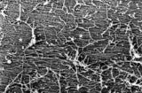 |
Surface of olfactory epithelium in the nose (rat) | Scanning electron microscopy. A carpet of fine long threads of olfactory cilia (*). On top of the carpet broken remnants of cilia clotted due to secretion products. | Olfactory epithelium | Poja Histology Collection - Respiratory System Subset |
| 277 |
 |
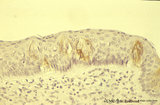
Surface of the nasal septum (rat) | Scanning electron microscopy of anterior part of the nasal septum. The superficial squamous cells of the stratified epithelium are large cells provided with small stubby microvilli. Cell borders are well indicated (↓). | Nasal vestibulum; Stratified epithelium | Poja Histology Collection - Respiratory System Subset |
| 278 |
 |
Surface of trachea epithelium (rat) | Scanning electronmicroscopy. There is an abundancy of cilia with interspersed protrusions (↓) of goblet cells due to apical secretion. | Pseudostratified epithelium | Poja Histology Collection - Respiratory System Subset |
| 279 |
 |
Survey frontal section of larynx (human) | Stain: Azan. A pseudostratified epithelium covers the mucosa of the laryngeal cavity and vestibulum. At the vocal fold edge (3) the epithelium appears to be nonkeratinizing squamous. The connective tissue is rigidly attached to the this edge and merges into the vocal ligament (dense elastic) and voc... | Vocal muscle; Vocal ligament; Laryngeal glands; Ventricular fold | Poja Histology Collection - Respiratory System Subset |
| 280 |
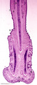 |
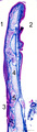
Survey nasal septum (dog) | Stain: Hematoxylin and eosin. The nasal septum consists of a cartilage skeleton present as one long (1, cartilago vomeronasalis) and two short plates (2, cartilago septi nasi) with centrally dark-stained areas of chondrocytes. Below, the distal tip is covered with keratinized stratified squamous epi... | Squamous stratified epithelium; Nasal vestibulum; Venous sinusoids; Venous plexus; Swell bodies | Poja Histology Collection - Respiratory System Subset |
| 281 |
 |
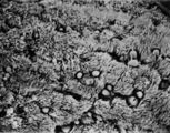
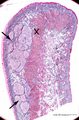
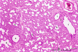
Survey of a centrilobular lung emphysema: section of an lobule (human, adult) | Stain: Hematoxylin and eosin. The enlargement of large areas of the air spaces (X) is evident due to destruction of the walls of alveoli throughout the lobule. | Poja Histology Collection - Respiratory System Subset | |
| 282 |
 |
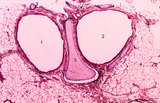
Survey of bronchus and lung parenchym (human, adult) | Stain: Azan. (1) Lumen of small bronchus beside a lumen of pulmonary artery (2). Thin plates of hyaline cartilage (3) and connective tissue surrounding (1) and (2). Arrows (→) indicate lung alveoli with black-stained patches of carbon deposits. (4) Lung parenchym with alveoli. | Lung parenchym | Poja Histology Collection - Respiratory System Subset |
| 283 |
 |
Survey of bronchus and lung parenchym (human, adult) | Stain: Azan. Lumina of bifurcated bronchi (1, 2). Thin plates of hyaline cartilage (3) and connective tissue surround the bronchi. Arrows (→) indicate lung alveoli with black-stained patches of carbon deposits. (4) Lung parenchyma with alveoli. | Lung parenchym | Poja Histology Collection - Respiratory System Subset |
| 284 |
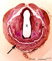 |
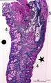
Survey of cross-section through larynx-esophagus (human) | Stain: Hematoxylin and bordeaux light. X = lumen of larynx (middle part); ↑ = esophagus (below) with an undulating mucosa covered with squamous epithelium. Elastic arythenoid cartilage (1); thyreoarythenoid muscle (2); thyroid cartilage (3); hyoid muscle (4). | Arythenoid cartilages | Poja Histology Collection - Respiratory System Subset |
| 285 |
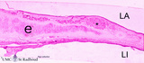 |
Survey of epiglottis (human) | Stain: Azan. Centrally light-stained elastic cartilage (e). Lingual side (LI) is covered with squamous epithelium. At the laryngeal side (LA) the squamous epithelium usually turns into respiratory epithelium with seromucous laryngeal glands (*). | Laryngeal gland; Oral cavity | Poja Histology Collection - Respiratory System Subset |
| 286 |
 |
Survey of epiglottis (human) | Stain: Azan. Centrally light-stained elastic cartilage (4). Lingual side (2) is covered with squamous epithelium. At the laryngeal side (1) the squamous epithelium usually turns into respiratory epithelium with seromucous laryngeal glands (5). Note also lymphoid aggregations in this area (3 →). | Oral cavity; Laryngeal glands | Poja Histology Collection - Respiratory System Subset |
| 287 |
 |
Survey of lip (human) | Stain: Azan. Arrows: labial glands in submucosa (mucous inner surface with non-keratinized epithelium). X: orbicularis oris muscle. Right side = skin surface (keratinized epithelium). Top = transitional zone (red zone) with slightly keratinized epithelium. | oral cavity; red zone; labial glands | Poja Histology Collection - Oral Cavity Subset |
| 288 |
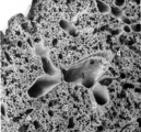 |
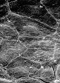
Survey of lung parenchym (gerbil) | Scanning electron microscopy. The photograph shows (1) bifurcation of several bronchi in the lung parenchym with alveoli (2). The long arrows indicate the route of alveolar ducts (4 ↕ length, and cross 4↑). Pulmonary vessels (3). | Lung parenchym; Pulmonary vessels | Poja Histology Collection - Respiratory System Subset |
| 289 |
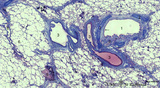 |
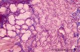
Survey of lung parenchym with bronchi and blood vessels (human, adult) | Stain: Azan. The bronchus (1) is surrounded with hyaline cartilage rings (2), seromucous glands (9) and muscle fibers (*), and lined with respiratory epithelium (←). Pulmonary arteries (3) in their neighborhood; a solitary pulmonary vein (8); (4) Carbon deposits of varying sizes (dust loaded macr... | Bronchiolus | Poja Histology Collection - Respiratory System Subset |
| 290 |
 |
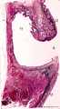
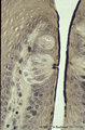
Survey of nose wing - human | Stain: Azan. At the top orofacial muscle (1, reddish) and hyaline cartilage (2, bluish) at the right. The external skin surface of the nose wing dot side (.) is covered with squamous epithelium, light-stained sebaceous glands (3) and hair structures (4). At the starred side (*) the nasal vestibulum... | Poja Histology Collection - Respiratory System Subset | |
| 291 |
 |
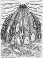
Survey of the nasal conchae (dog, isolated turbinate bones) | Stain: Hematoxylin and eosin. The conchae (turbinates) consist of three parts: the inferior, middle and superior turbinate bones (3, black-stained); they are covered by a respiratory mucosa (1) or by an olfactory mucosa (2). In humans only a small part of the superior concha exhibits olfactory epith... | Conchae nasales; Olfactory epithelium; Respiratory epithelium | Poja Histology Collection - Respiratory System Subset |
| 292 |
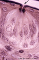 |
Taste bud of a foliate papil of the tongue (dorsal side, rabbit; high magnification) | Stain: Heidenhain's iron hematein. Taste bud with slender cells and darker stained spiky nuclei (type I cell), and cells with light stained round nuclei (type II cells). Below axons of the gustatory nerve fibers and nuclei of Schwann cells. Arrow points to narrow pore where dark stained fluffy struc... | oral cavity; foliate papillae; taste pore | Poja Histology Collection - Oral Cavity Subset |
| 293 |
 |
Taste bud of a foliate papilla of the tongue (dorsal side, human) | Stain: antikeratin-7 / immunoperoxidase and hematoxylin counterstained. Type I cells with small slender nuclei are identified by positive staining with cytokeratin 7 (monoclonal antibody OVTL12/30). | cytokeratin; oral cavity; foliate papilla; von Ebner | Poja Histology Collection - Oral Cavity Subset |
| 294 |
 |
Taste bud of the tongue | Scheme electronmicroscopy. Generally a taste bud is composed of about 20 to 70 spindle-shaped epithelial cells. Slender type I cell ('Dark' cell) and oval shaped type II cell ('Light' cell) with a pale, round nucleus with their apices end in microvilli in the taste pore. The number of type I cells i... | oral cavity; foliate papillae; taste pore; electronmicroscopy | Poja Histology Collection - Oral Cavity Subset |
| 295 |
 |
Taste buds of foliate papillae of the tongue (dorsal side, rabbit) | Stain: Heidenhain's iron hematein. Taste buds within the lining (non-keratinized) epithelium of the groove. Note the pore in the left taste bud, with darkly stained fluffy apical microvilli. The lightly stained gustatory nerve fibers (nuclei of Schwann cells) approach the left taste buds. The taste ... | oral cavity; foliate papillae; taste pore | Poja Histology Collection - Oral Cavity Subset |
| 296 |
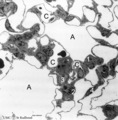 |
The terminal part of the airway: alveoli (dog) | Electron microscopy (low magnification). (A) indicate alveoli; (C) indicate capillaries. (1) type I alveolar cell (pneumocyte I, squamous alveolar cell); (2) type II alveolar cell (pneumocyte II, great alveolar cell). (3) alveolar macrophage. (4) endothelium of capillary. | Pneumocyte I; Pneumocyte II; Alveolar macrophage | Poja Histology Collection - Respiratory System Subset |
| 297 |
 |
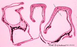
Terminal sac period of developing lung (human, fetus) | Stain: Hematoxylin and eosin. The transition of a terminal bronchiolus (1) into two future respiratory bronchioli (2), present as dilated spaces (saccules derived from the primitive respiratory channels, hence the name terminal sac period). The surrounding cellular tissue is composed of developing p... | Lung development; Terminal sac period; Respiratory bronchioli; Mesenchyme | Poja Histology Collection - Respiratory System Subset |
| 298 |
 |
Terminal sac period of developing lung (human, fetus, low magnification) | Stain: Hematoxylin and eosin. At the right cross-sections of a large bronchus (1) with cartilage rings (2). Close to them a pulmonary artery (3), and a bronchiolus (4) without cartilage. (5) indicates a respiratory bronchiolus. Most alveoli are not inflated, the numerous dilated spaces (6) are saccu... | Lung development ; Terminal sac period; Respiratory bronchioli | Poja Histology Collection - Respiratory System Subset |
| 299 |
 |
Tongue (lingual gland, human) | Stain: Azan. The posterior lingual gland is composed of areas with pure serous acini neighbouring pure mucous acini between the tongue muscles (at the left few striated fibers). | oral cavity; lingual gland | Poja Histology Collection - Oral Cavity Subset |
| 300 |
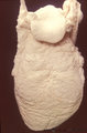 |
Tongue (macroscopy, dorsal side, human) | At the bottom (apex) of the picture the dorsal side is covered with numerous closed packed, small, conical filiform papillae. Interspersed among them are the larger fungiform papillae. Posteriorily the circumvallatae papillae are arranged in a V-shape row. Behind the V-row the area is covered with l... | oral cavity; papillae | Poja Histology Collection - Oral Cavity Subset |