The Health Education Assets Library (HEAL) is a collection of over 22,000 freely available digital materials for health sciences education. The collection is now housed at the University of Utah J. Willard Marriott Digital Library.
TO
Filters: Collection: "ehsl_heal" Format: image
| Title | Description | Subject | Collection | ||
|---|---|---|---|---|---|
| 276 |
 |
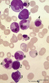
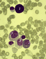
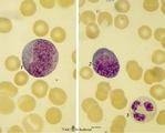
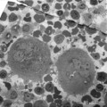
Multiple myeloma, plasma cell leukemia in bone marrow smear (human) | Stain: May-Grnwald-Giemsa (MGG). Mutiple myeloma (Kahler's disease) is characterized by proliferation of abnormal plasma cells (1, myeloma cells) in the bone marrow. In the great majority of patients secretion of a single homogenous immunoglobulin product (monoclonal component) occurs. Note the ecce... | Poja Histology Collection - Blood & Bone Marrow Subset | |
| 277 |
 |
Pleiomorph normal bone marrow smear (human) | Stain: May-Grnwald-Giemsa (MGG). The normal bone marrow with pleiomorphism, i.e. contains all stages of the erythroblastic differentiation series (1-5) as well as the stages of the myeloblastic series (6). (1) Basophilic erythroblast. (2),(3),(4) Polychromatic erythroblasts. (5) Orthochromatic er... | Poja Histology Collection - Blood & Bone Marrow Subset | |
| 278 |
 |
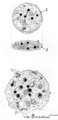
Lymphocyte | Scheme electron microscopy. A lymphocyte with an indented nucleus and (1) nucleolus shows a juxta-nuclear Golgi area (2) and centriole (3), several mitochondria (4) and sparsely profiles of rough endoplasmic reticulum (5) in the cytoplasm. Few dense-stained granules (6) are present. The granules con... | Poja Histology Collection - Blood & Bone Marrow Subset | |
| 279 |
 |
Small, medium and large lymphocytes in blood smear (human) | Stain: May-Grnwald-Giemsa (MGG). The picture shows three activation stages of lymphocytes as (1) small, (2) medium and (3) large lymphocytes. In that range the nucleus becomes less condensed and more transparent, while the amount of cytoplasm increases. The large lymphocyte also contains some granul... | Poja Histology Collection - Blood & Bone Marrow Subset | |
| 280 |
 |
Myeloid cells in bone marrow smear (human) | Stain: May-Grnwald-Giemsa (MGG). (1) promyelocyte is the largest cell in the myeloid series. It has a transparent nucleus with nucleoli and ample cytoplasm with many azurophilic granules. (2) myelocyte. (3) metamyelocyte with an indented nucleus. (4) beginning of nucleus segmentation in the band neu... | Poja Histology Collection - Blood & Bone Marrow Subset | |
| 281 |
 |
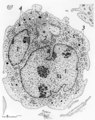
Erythroblastic island in bone marrow | Scheme electron microscopy. Different stages of maturing erythroblasts (2) are exposed in this scheme. The centrally localized phagocytic reticular cell (1) has many long cytoplasmic extensions that form a network with similar cells within the bone marrow. Its nucleus is irregular. The cytoplasm has... | Poja Histology Collection - Blood & Bone Marrow Subset | |
| 282 |
 |
Peroxidase activity in neutrophilic granulocyte (peripheral blood, guinea pig) | Electron microscopy (peroxidase reaction with diaminobenzidin staining). The elongated nucleus (3) of this strongly ameboid phagocytic cell has several projections. These nuclear extensions are interconnected by thin heterochromatin strands. The cytoplasm exhibits moderate amounts of organelles, gly... | Poja Histology Collection - Blood & Bone Marrow Subset | |
| 283 |
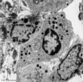 |
Plasma cells with Russell bodies (nose septum, rat) | Electron microscopy. Accumulation of few plasma cells. These mature plasma cells exhibit in their cytoplasm's dilated rough endoplasmic reticula (1) filled with electron-grey material (antibodies) as well as electron-dense granules (3) of varying sizes confirming their secretory activities. (2) indi... | Poja Histology Collection - Blood & Bone Marrow Subset | |
| 284 |
 |
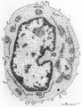
Pro-megakaryocyte | Scheme electron microscopy. The pro-megakaryocyte (20-80 m) develops from a basophilic megakaryoblast (up to 50 m) that originates from a committed stem cell. The large pro-megakaryocyte starts with the early synthesis of the characteristic granules of platelets. Apart from a bilobed nucleus (1) and... | Blood; Bone Marrow; Electron microscopy; Pro-megakaryocyte; Megakaryocyte | Poja Histology Collection - Blood & Bone Marrow Subset |
| 285 |
 |
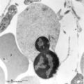
A bare megakaryocyte nucleus and myelocytes in bone marrow smear (human) | Stain: May-Grnwald-Giemsa (MGG). (1) Indicates a bare large-sized polyploid nucleus of a megakaryocyte which has totally shed the cytoplasm. These cells are often seen in normal marrow and are ultimately removed by macrophages. There is no cytoplasm left, nor platelets are left around the nucleus. T... | Poja Histology Collection - Blood & Bone Marrow Subset | |
| 286 |
 |
Promyelocyte and basophilic erythroblast in bone marrow smear (human) | Stain: May-Grnwald-Giemsa (MGG). The composition shows a (pro)myelocyte (1) along a basophilic erythroblast (2). The (pro)myelocyte nucleus is less condensed, contains nucleoli and the light basophilic cytoplasm is filled with azurophilic granules. The cytoplasm of the erythroblast is strong basophi... | Poja Histology Collection - Blood & Bone Marrow Subset | |
| 287 |
 |
Myeloblast in bone marrow smear (human) | Stain: May-Grnwald-Giemsa (MGG). The myeloblast (1) shows a very transparent nucleus and several distinct nucleoli. The slightly basophilic cytoplasm is limited to a small rim in contrast to a promyelocyte. No granules are yet visible. (2) small lymphocyte. | Blood; Bone Marrow; Myeloblast; Lymphocyte | Poja Histology Collection - Blood & Bone Marrow Subset |
| 288 |
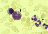 |
Promonocyte in bone marrow smear (human) | Stain: May-Grnwald-Giemsa (MGG). The promonocyte (1) has an irregular nucleus and shows the first step towards indentation (arrow). The cell has a characteristic greyish-blue cytoplasm. (2) Represents three young neutrophils. (3) A plasmacytoid lymphocyte. | Poja Histology Collection - Blood & Bone Marrow Subset | |
| 289 |
 |
Reactive promyelocyte during sepsis or chemotherapy in peripheral bood smear (human) | Stain: May-Grnwald-Giemsa (MGG). Neutrophilic promyelocyte with toxic granulation and vacuolar degeneration. Note that nucleoli are still visible. | Poja Histology Collection - Blood & Bone Marrow Subset | |
| 290 |
 |
Platelets | Scheme electron microscopy. These disc-shaped anucleate cells (2-4 m) are derived from cytoplasmic fragments of the megakaryocyte. They contain few mitochondria, a canalicular system, a smooth tubular system, marginal localized microtubules, glycogen and different types of granula. A circumferential... | Poja Histology Collection - Blood & Bone Marrow Subset | |
| 291 |
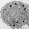
 |
Monocyte (peripheral blood, human) | Electron microscopy. In this picture the large nucleus of this cell (diameter of 12-20 μm) is twice sectioned (1). Golgi areas (2), a few profiles of rough endoplasmic reticulum (3) and many free ribosomes are present. There are many mitochondria (4) as well as small-sized light vesicles (5). The c... | Poja Histology Collection - Blood & Bone Marrow Subset | |
| 292 |
 |
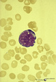
A plasmacytoid lymphocyte in peripheral blood smear (human) | Stain: May-Grnwald-Giemsa (MGG). The plasmacytoid lymphocyte is an activated B lymphocyte to be transformed into a plasma cell. The cytoplasm is more basophilic and the chromatin pattern is more clumped than in a virgin small lymphocyte. When the cell contains numerous immunoglobulin inclusions (glo... | Poja Histology Collection - Blood & Bone Marrow Subset | |
| 293 |
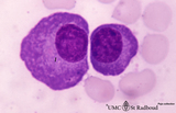 |
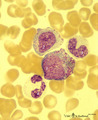
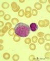
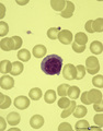
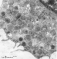
Plasma cells in bone marrow smear (human) | Stain: May-Grnwald-Giemsa (MGG). The two plasma cells are mature antibody-producing cells. The basophilic cytoplasm is filled up with rough endoplasmic reticulum (RER). The lighter zone (1) represents the Golgi areas. The nucleus is situated eccentrically and contains strands of condensed chromatin ... | Poja Histology Collection - Blood & Bone Marrow Subset | |
| 294 |
 |
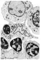
Survey of megakaryocytes (peripheral blood, human) | Electron microscopy. In a section of a so-called buffy coat collected from peripheral blood two huge polyploid megakaryocytes (1) (diameter up to 160 μm) are present. Note the dark granules in the future platelets. (2) erythroblasts; (3) reticulocytes and erythrocytes; (4, 5) monocytes. The arrows ... | Poja Histology Collection - Blood & Bone Marrow Subset | |
| 295 |
 |
Orthochromatic erythroblast (bone marrow, rabbit) | Electron microscopy. The orthochromatic erythroblast (1) or late normoblast shows the early stage of extrusion of the nucleus though the latter appears not yet fully pyknotic. The cytoplasm contains few slender mitochondria (2) as well as scattered clumped polysomes. (3) Part of a reticulocyte. | Poja Histology Collection - Blood & Bone Marrow Subset | |
| 296 |
 |
Survey of bone marrow section (human) | Stain: modified hematoxylin and eosin. In this survey the white spots (1) are fat cells. The marrow is well filled with cells of erythropoietic and myelopoietic series. The very large cells are usually megakaryocytes. Cells with very dark condensed nuclei belong mostly to the erythropoietic series. | Poja Histology Collection - Blood & Bone Marrow Subset | |
| 297 |
 |
Megakaryoblast in bone marrow smear (human) | Stain: May-Grnwald-Giemsa (MGG). The megakaryoblast (1) is not yet producing platelets and has a relative small amount of basophilic cytoplasm. The nucleus is large with fine disperse chromatin and nucleoli. Most other nucleated cells are of the myeloid cell line. | Poja Histology Collection - Blood & Bone Marrow Subset | |
| 298 |
 |
Erythroblast and neutrophilic myelocytes in bone marrow smear (human) | Stain: May-Grnwald-Giemsa (MGG). (1) an older basophilic erythroblast with slightly condensed nuclear chromatin. (2) two neutrophilic myelocytes with azurophilic primary granules. | Poja Histology Collection - Blood & Bone Marrow Subset | |
| 299 |
 |
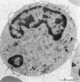
Band form of neutrophilic granulocyte | Scheme electron microscopy. From CFU-S (colony forming units-spleen) stem cells arise CFU-GM (colony forming unit-granulocyte/monocyte) stem cells. The latter divide by mitoses and differentiate via promyeloblasts and myeloblasts into neutrophilic myelocytes (the last proliferative stage). The next ... | Poja Histology Collection - Blood & Bone Marrow Subset | |
| 300 |
 |
Mast cell (lung, human) | Electron microscopy. Mast cells (mastocytes) are frequently found perivascularly or perineurally. A part of this mast cell shows thin microvilli at the surface and the cytoplasm is provided with a moderate amount of organelles. At (*) the Golgi area (1) is seen in close association with granules tha... | Poja Histology Collection - Blood & Bone Marrow Subset |