The Health Education Assets Library (HEAL) is a collection of over 22,000 freely available digital materials for health sciences education. The collection is now housed at the University of Utah J. Willard Marriott Digital Library.
TO
Filters: Collection: ehsl_heal
| Title | Description | Subject | Collection | ||
|---|---|---|---|---|---|
| 251 |
 |
Lip (human), mucoserous labial glands in mucous inner surface | Stain: Azan. Mixed labial glands with serous demilunes (von Ebner-Giannuzzi), a few myoepithelial cells, and a striated duct (right upper corner). | oral cavity; lining mucosa; labial glands | Poja Histology Collection - Oral Cavity Subset |
| 252 |
 |
Lip (human), mucous inner surface | Stain: Azan. Non-keratinized epithelium with high, narrow dermal papillae and many capillaries. Mixed labial glands (seromucous) and few skeletal muscle fibers (orbicularis oris) in the submucosa. | oral cavity; lining mucosa; labial glands | Poja Histology Collection - Oral Cavity Subset |
| 253 |
 |
Lip (human), mucous inner surface | Stain: Azan. Non-keratinized epithelium with high, narrow dermal papillae and many capillaries. Note lymphocytic infiltrates and the capillaries in the dermal papillae. | oral cavity; lining mucosa | Poja Histology Collection - Oral Cavity Subset |
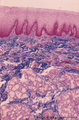
| 254 |
 |
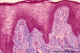
Lip (human), outer (left) and inner side (right) | Stain: Azan. Left image: keratinized squamous epithelium with thin red cornified layer, hair follicle, sebaceous glands and skeletal muscle fibers (orbicularis oris). Right image: non-keratinized epithelium with high, narrow dermal papillae and more capillaries. Mixed labial glands (seromucous), f... | oral cavity; lining mucosa | Poja Histology Collection - Oral Cavity Subset |
| 255 |
 |
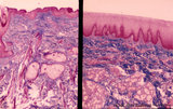
Lip (human), region between red zone (vermilion border) and mucosa inner surface | Stain: Azan. Bundles of skeletal muscle fibers (musculus orbicularis oris), highly vascularized lamina propria. Note the narrow dermal papillae on the left side (inner lip) and the broad irregular papillae on the right side (red zone of the lip). | oral cavity; lining mucosa; red zone; vermilion border | Poja Histology Collection - Oral Cavity Subset |
| 256 |
 |
Lip (human), skin surface. | Stain: Azan. Keratinized squamous epithelium with hair follicles; submucosa with sebaceous glands, and extensions of skeletal muscle cells (orbicularis oris). | oral cavity | Poja Histology Collection - Oral Cavity Subset |
| 257 |
 |
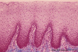
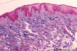
Lip (human), transitional zone (red zone or vermilion border) | Stain: Azan. Slightly cornified epithelium with high irregular dermal papillae and many capillaries. Note the epithelium is thicker, but less cornified than the epidermis. The red color of the lips is due to the rich vascularity of the lamina propria and the lucidity of the epithelium. | oral cavity; lining mucosa; red zone; vermilion border | Poja Histology Collection - Oral Cavity Subset |
| 258 |
 |
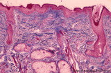
Lip (human), transitional zone (red zone or vermilion border) | Stain: Azan. Slightly cornified epithelium with high irregular dermal papillae and many capillaries. Fat cells and skeletal muscle cells are located in the submucosa. | oral cavity; lining mucosa; red zone; vermilion border | Poja Histology Collection - Oral Cavity Subset |
| 259 |
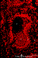 |
Localization of B cells in spleen (rat) | Stain: Immunofluorescence of Vector red using Mark-1 antibody against B cells. The T cells in the PALS (periarteriolar lymphatic sheath) remain unstained (1, dark). The red-stained B cells are packed in the germinal centre (2), the corona (3) or mantle layer that diffuses into the marginal zone and... | B lymphocytes; immunofluorescence; PALS | Poja Histology Collection - Lymphatic Tissues and Organs Subset |
| 260 |
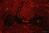 |
Localization of heparan sulfate (HS) in spleen (rat) | Stain: Immunofluorescence of Alexa 594 red labeled single chain antibody 3C3 for heparan sulfate (HS). The antibody stains HS epitopes of the meshwork of reticulum cells and the basal membrane of blood vessels. (1) central arteries in the T cell area of the PALS (2). The B cell area in the follic... | white pulp; heparan sulfate; PALS | Poja Histology Collection - Lymphatic Tissues and Organs Subset |
| 261 |
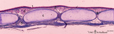 |
Longitudinal section of trachea (human, adult) | Stain: Azan. On top: the epithelium (1) is pseudostratified with cilia and goblet cells. The dense lamina propria (2, ↓, deep blue) is demarcated from the submucosa with seromucous tracheal glands (3). C-shaped cartilage rings (C, light blue) maintain patency. The cartilage edges are connected by ... | Pseudostratified epithelium ; Seromucous glands; Tracheal glands; Fibroelastic membrane ; Adventitia | Poja Histology Collection - Respiratory System Subset |
| 262 |
 |
Longitudinal section of trachea (human, adult) | Stain: Azan. On top: the epithelium (1) is pseudostratified with cilia and goblet cells on a distinct basement membrane (↓). There is almost no demarcation (at this magnification) between lamina propria and submucosa due to the dense blue (2) staining of elastic fibers reinforced by collagen fiber... | Pseudostratified epithelium ; Tracheal glands; Fibroelastic membrane | Poja Histology Collection - Respiratory System Subset |
| 263 |
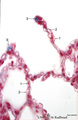 |
Lung alveoli (human) | Stain: Azan. Alveoli (diameter about 200 m) are formed by thin walls of epithelial cells (1) such as type I alveolar cells and (blue stained) fine collagen (3) and elastin intermingled with dilated capillaries (2). | Poja Histology Collection - Respiratory System Subset | |
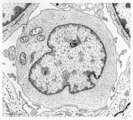
| 264 |
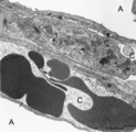 |
Lung capillaries and air-blood barrier (rat) | Electron microscopy. Two neighboring capillaries (C) and the alveolar spaces (A). The endothelial cell (1) of the upper capillary contains an organelle-rich cytoplasm, a centriole (2), contractile filaments (↓) and electron-dense membrane-bound granules, the so-called Weibel-Palade bodies. These b... | Poja Histology Collection - Respiratory System Subset | |
| 265 |
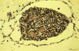 |
Lymph node (fetus, human) | Stain: Trichrome (Goldner). Along the course of lymphatic vessels lymph nodes develop. Lymphatic vessels dilate and form lymphatic sacs and lymphocytes aggregate around these sacs. A tiny developing lymph node with dense-stained thymocyte nuclei is closely associated with dilated lymph capillaries. ... | fetal lymph node; development | Poja Histology Collection - Lymphatic Tissues and Organs Subset |
| 266 |
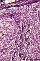 |
Lymph node (human) | Stain: Silver stain (Gomori). Due to the argyrophilia of reticular fibres the reticular network is black-stained among others such as collagen type I, III, IV. From the fibrous capsule (1) a meshwork of fine reticular fibres penetrates into the cortex of a lymph node crossing the subcapsular (or m... | germinal center; argyrophilia; reticular fibers | Poja Histology Collection - Lymphatic Tissues and Organs Subset |
| 267 |
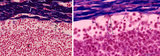 |
Lymph node (human) | Stain: Azan. Left: subcapsular (or marginal) sinus in lymph node. Right: higher magnification. Both pictures show a thick capsule (1), at (2) subcapsular (or marginal) sinus filled with lymphocytes indicate (lining) littoral cells (?). Reticular cells (3) are localized perpendicularly through sin... | subcapsular sinus; reticular cell | Poja Histology Collection - Lymphatic Tissues and Organs Subset |
| 268 |
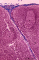 |
Lymph node (human) | Stain: Azan. Part of the cortex of a lymph node with the capsule (1) and subcapsular (or marginal) sinus (2) filled with lymphocytes. From the blue-stained dense capsule a long trabecula (3) flanked by paratrabecular (intermediate) sinuses penetrates between the paracortical areas (6). Paratrabecula... | Poja Histology Collection - Lymphatic Tissues and Organs Subset | |
| 269 |
 |
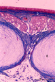
Lymph node (human) | Stain: Azan. Part of the cortex of a lymph node with the capsule (1) and subcapsular (or marginal) sinus (2) filled with lymphocytes. Below the subcapsular sinus a darkly stained rim or corona (3) of small lymphocytes (B cells) surrounds the lucid stained germinal centre (4). The white spaces repre... | follicle; germinal center | Poja Histology Collection - Lymphatic Tissues and Organs Subset |
| 270 |
 |
Lymph node (human) | Stain: Azan. Left and right: survey of lymph nodes, compare the individual differences between the nodes. The pictures display the cortex (1) with capsule (3), and the medulla (2). The cortex consists of (secondary) follicles displaying a clear germinal centre (4). The paracortex (T cell area) is ... | follicle; paracortex; cortex; germinal center | Poja Histology Collection - Lymphatic Tissues and Organs Subset |
| 271 |
 |
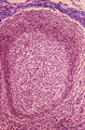
Lymph node (mouse) | Stain: Hematoxylin & eosin. For comparison with the human lymph node a survey of a small rodent lymph node is demonstrated. Basically the same divisions are found: (1) a cortex (outer cortex zone, nodular cortex or superficial cortex) covered with a (2) thin capsule and subcapsular sinus; a paracor... | follicle; hilum; medulla; cortex | Poja Histology Collection - Lymphatic Tissues and Organs Subset |
| 272 |
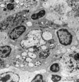 |
Lymph node (rat) | Electron microscopy. In the paracortical area (inner cortex) of a lymph node interdigitating cells (1) are found (so-called IDC, antigen-presenting cell or APC). They show branched extensions (3) in between the closely apposed surrounding T lymphocytes (2). In the cytoplasm of the IDC lysosomal inc... | electron microscopy; interdigitating cell; antigen-presenting cell | Poja Histology Collection - Lymphatic Tissues and Organs Subset |
| 273 |
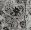 |
Lymph node (rat) | Electron microscopy. Within the reticular framework of lymph nodes a variety of active reticular cells are present, some of them (1) have phagocytised foreign material (1a) and contain numerous lysosomal structures (1a) and large Golgi areas (1b)). Others are less active (2). (3) wandering interdig... | electron microscopy; interdigitating cell; antigen-presenting cell | Poja Histology Collection - Lymphatic Tissues and Organs Subset |
| 274 |
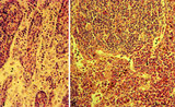 |
Lymph nodes (rat) | Stain: Hematoxylin & pyronin. Pyronin was formerly used to demonstrate young blast cell types rich in polysomes and packed rough endoplasmic reticulum (RER) resulting in a reddish stain of the cytoplasm. A: Part of the medulla with medullary cords (1) and medullary sinuses (2) close to the hilum o... | infection; medullar cord; plasma cells ; macrophages | Poja Histology Collection - Lymphatic Tissues and Organs Subset |
| 275 |
 |
Lymphoblast | Scheme electron microscopy. The lymphoblast shows a high nucleus-cytoplasm ratio, it is a large cell (10-20 μm) with a large nucleus and distinct nucleolus. Apart from numerous ribosomes, a small juxta-nuclear Golgi area the cytoplasm is scanty with very few organelles. | Poja Histology Collection - Blood & Bone Marrow Subset |