The Health Education Assets Library (HEAL) is a collection of over 22,000 freely available digital materials for health sciences education. The collection is now housed at the University of Utah J. Willard Marriott Digital Library.
Filters: Collection: ehsl_heal
| Title | Description | Subject | Collection | ||
|---|---|---|---|---|---|
| 251 |
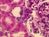 |
Kidney | At the vascular pole of this renal corpuscle can be seen cross-sections of the afferent and efferent arterioles. A well-defined macula densa is present in the wall of the distal convoluted tubule (DCT), adjacent to the afferent arteriole. There are also a number of cross-sections of proximal convolu... | Kidney; Renal Corpuscle | UCLA Histology |
| 252 |
 |
Kidney | In this image we see the glomerulus of a renal corpuscle, surrounded by dark red proximal convoluted tubules (PCT) and lighter stained distal convoluted tubules (DCT). The basement membranes of the parietal wall of Bowman's capsule and surrounding tubules are stained blue. UCLA Histology Collection. | glomerulus; Kidney; trichrome | UCLA Histology |
| 253 |
 |
Kidney | In this higher power view of the above image, the wall of the minor calyx can be seen to consist of transitional epithelium. The bands of smooth muscle and connective tissue of the wall of the calyx can be seen clearly. UCLA Histology Collection. | Kidney; Minor Calyces; Minor Calyx | UCLA Histology |
| 254 |
 |
Kidney | A number of features are seen in this glomerulus. Identify the simple squamous epithelium of the parietal layer of the renal corpuscle. Also note the dark podocyte nuclei of the visceral layer which wrap around the many glomerular capillaries. UCLA Histology Collection. | glomerulus; Kidney; renal corpuscle | UCLA Histology |
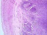
| 255 |
 |
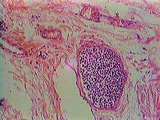
Lymph Node | Convex surface of a bronchial lymph node showing the rather thick connective tissue capsule and subcapsular sinus through which lymph enters the lymph node. Phagocytic macrophages are present and have engulfed black particulate matter such as carbon or dust. Cortical sinuses, through which lymph per... | Lymph Node; macrophage; subcapsular sinus | UCLA Histology |
| 256 |
 |
Lymph Nodes | This image from the parenchyma of the lymph node shows phagocytic macrophages crammed with carbon. Few lymphocytes are visible. The connective tissue stroma contains reticular fibers, fibroblasts, and reticular cells. UCLA Histology Collection. | Lymph Node; lymphocytes; macrophage | UCLA Histology |
| 257 |
 |
Lymph Nodes | This silver stained slide reveals the collagen and reticular fibers of the lymph node. Note the thick connective tissue capsule and its extension, the trabecula. Directly under the capsule is the subcapsular sinus that receives lymph flowing in from afferent lymphatic vessels. From the subcapsular s... | Lymph Node; silver stain; subcapsular sinus | UCLA Histology |
| 258 |
 |
Lymph Nodes | This section demonstrates medullary cords that contain packed strips of lymphocytes. Between the cords are spaces known as medullary sinuses through which lymph percolates. The cortex of this lymph node contains secondary follicles or nodules, which show pale central regions, the germinal centers. N... | germinal center; Lymph Node; medullary cords; medullary sinuses | UCLA Histology |
| 259 |
 |
Lymph Nodes | This image shows the hilum of the lymph node, from which efferent lymphatic vessels exit. Arteries and veins (not visible) also enter and exit here. Note vestiges of cortex with nodules containing germinal centers. Strips of medullary cords can also be seen. UCLA Histology Collection. | germinal center; hilum; Lymph Node | UCLA Histology |
| 260 |
 |
Lymph Nodes | Identify the capsule, the underlying subcapsular sinuses, and the trabecula. At the convex surface of the lymph node, afferent lymphatic vessels enter the node. Note numerous lymphocytes in the cortex. UCLA Histology Collection. | Lymph Node; lymphocytes; trabecula | UCLA Histology |
| 261 |
 |
Lymph Nodes | This image shows a cross section of an efferent lymphatic vessel in the hilar region. Like veins, lymphatic vessels are thin-walled. Unlike veins, lymphatic vessels are filled with lymphocytes. The hilum contains large amounts of collagen fibers. UCLA Histology Collection. | Lymph Node; lymphatic vessel | UCLA Histology |
| 262 |
|
Lymph Nodes | This section demonstrates medullary cords that contain packed strips of lymphocytes. Between the cords are spaces known as medullary sinuses through which lymph percolates. The cortex of this lymph node contains secondary follicles or nodules, which show pale central regions, the germinal centers. N... | germinal center; Lymph Node; medullary cords; medullary sinuses | UCLA Histology |
| 263 |
 |
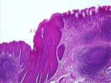
lymphoid tissue | This image shows stratified squamous epithelium overlying connective tissue. The parenchyma contains many lymphoid nodules. Crypts are present where epithelium invaginates into parenchyma. UCLA Histology Collection. | lymphoid nodule; lymphoid tissue; palatine tissue; palatine tonsil | UCLA Histology |
| 264 |
 |
Lymphoid Tissue | The thick stratified squamous epithelium of the esophagus changes into thin simple columnar epithelium of the stomach. In the esophageal region, note two lymphoid nodules with clear germinal centers. Diffuse lymphoid tissue is found scattered throughout the inner layers of the digestive tract. UCLA ... | Esophagus; germinal center; Lymphoid Tissue; Stomach | UCLA Histology |
| 265 |
 |
Lymphoid Tissue | Esophageal region; stratified squamous epithelium changes into thin columnar epithelium of stomach. In esophageal region, note lymphoid nodules with clear germinal centers. Diffuse lymphoid tissue is scattered throughout submucosa. UCLA Histology Collection. | Esophagus; germinal center; lymphoid nodule; Stomach | UCLA Histology |
| 266 |
 |
Lymphoid Tissue | At a higher magnification, identify Brunner's glands and the lymphoid nodule with germinal center. UCLA Histology Collection. | Brunner's glands; Lymphoid tissue; Small Intestine | UCLA Histology |
| 267 |
 |
Lymphoid Tissue | The simple columnar epithelium of the ileum is thrown into villi. Crypts of Lieberkuhn are found between the bases of villi. Mucosal aggregations of lymphoid nodules, known as Peyer's Patches, can be seen here. Some contain germinal centers. UCLA Histology Collection. | Lymphoid Tissue; Peyer's Patches; Small intestine | UCLA Histology |
| 268 |
 |
Lymphoid Tissue | The lumen of the appendix communicates with crypts of Lieberkuhn. Lymphoid nodules of various size can be found here, along with germinal centers. The outer smooth muscle layer, or muscularis externa, is visible at right. UCLA Histology Collection. | Appendix; Large intestine; Lymphoid Tissue; with germinal centers | UCLA Histology |
| 269 |
 |
lymphoid tissue | The gastric (stomach) region is lined by simple columnar epithelium. Note diffuse lymphoid tissue. On the right, identify the lymph nodule and its pale germinal center. UCLA Histology Collection. | germinal center; lymph nodule; lymphoid tissue; Stomach | UCLA Histology |
| 270 |
 |
Lymphoid Tissue | Diffuse lymphoid tissue contains numerous lymphocytes not organized into discrete nodules. Lymphocytes are interspersed in the mucosa which contains red stained parietal cells. UCLA Histology Collection. | lymphocytes; lymphoid tissue; nodules; Stomach | UCLA Histology |
| 271 |
 |
Male Reproductive System | The seminal vesicles are long coiled tubes with a distinct lamina propria. Note the convoluted nature of the vesicles lumen. The mucosa is highly folded, and the muscular coat is composed of two layers of smooth muscle - an inner circular layer, and a outer longitudinal layer. UCLA Histology Collect... | male reproductive system; Seminal vesicle | UCLA Histology |
| 272 |
 |
Male reproductive system | A seminiferous tubule showing a Sertoli cell with a pale staining nucleus and a prominent nucleolus. Spermatogonia lie along the basal lamina and are either dark or more pale staining. After division the daughter cells of the spermatogonia, the primary spermatocytes work their way toward the lumen a... | Male reproductive system; Seminiferous tubule; Sertoli cell; testis | UCLA Histology |
| 273 |
 |
male reproductive system | The bulk of the testis is formed by the seminiferous tubules. Please note that the tubules are surrounded by a basal lamina and stromal connective tissue. Within the seminiferous tubule, spermatogonia divide and mature to produce spermatozoa Within the surrounding stroma adipocytes, fibroblasts, and... | male reproductive system; Seminiferous tubule; testis; cells of Leydig | UCLA Histology |
| 274 |
 |
Male reproductive system | Higher power magnification of the transition from seminiferous tubule (lined with Sertoli cells, spermatogonia, spermatocytes, and spermatids) to the tubuli recti (lined with Sertoli cells) to the rete testis (lined with simple columnar cells). Note the dense, collagenous, connective tissue of the m... | male reproductive system; Seminiferous tubule; testis | UCLA Histology |
| 275 |
 |
Male Reproductive System | Erectile tissue of the corpus cavernosa consisting of connective tissue and vascular sinuses. UCLA Histology Collection. | corpus cavernosa; Erectile tissue; male reproductive system; Penis | UCLA Histology |