The Health Education Assets Library (HEAL) is a collection of over 22,000 freely available digital materials for health sciences education. The collection is now housed at the University of Utah J. Willard Marriott Digital Library.
TO
Filters: Collection: ehsl_heal
| Title | Description | Subject | Collection | ||
|---|---|---|---|---|---|
| 251 |
 |
Scheme of the epiglottis (human, adult) | The laryngeal side of the epiglottis (1) is covered with respiratory epithelium, while the lingual side (2) is similar to the oral cavity epithelium (squamous type). The transitional zone to respiratory epithelium is marked by (3). The scaffold consists of elastic cartilage (4). Seromucous laryngea... | Laryngeal glands; Oral cavity | Poja Histology Collection - Respiratory System Subset |
| 252 |
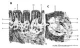
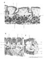 |
Scheme of tongue papillae and foliate papillae | A. Circumvallate papillae (tongue, human, adult) magnification x 35 objective. B. Foliate papillae (tongue, rabbit) magnification x 85 objective. C. Taste bud in foliate papilla (tongue, rabbit) magnification x 750 objective. 1. (circum)vallate papilla; 2. non-keratinized stratified squamous epit... | oral cavity; von Ebner; circumvallate papillae; foliate papillae | Poja Histology Collection - Oral Cavity Subset |
| 253 |
 |
Scheme of tongue papillae and taste buds | B. Scheme of foliate papillae of the tongue (rabbit) magnification x 85 objective. C. Scheme of taste bud in foliate papilla of the tongue (rabbit). 2. non-keratinized stratified squamous epithelium; 3. taste bud; 4. serous gland (von Ebner); 5. draining duct (of serous gland); 7. foliate papil... | oral cavity; von Ebner; foliate papillae | Poja Histology Collection - Oral Cavity Subset |
| 254 |
 |
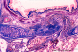
Scheme of tooth development (human) | A. Survey of tooth germ (bell stage, embryo) magnification x 35 objective. B. Formation of enamel and dentin (bell stage, embryo) magnification x 350 objective. 1. pulp organ; 2. odontoblasts; 3. mantle predentin; 4. dentin; 5. enamel; 6. initial enamel; 7. inner dental epithelium (ameloblasts);... | oral cavity; dental lamina; bell stage | Poja Histology Collection - Oral Cavity Subset |
| 255 |
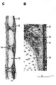 |
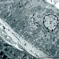
Scheme of trachea | C. Trachea (human, adult) magnification x 7.5 objective). D. Trachea (human, adult, detail of rectangle in C) magnification x 85 objective. (12) pseudostratified ciliated epithelium; (13) seromucous tracheal glands; (14) condensed layer of elastic fibers at the border of lamina propria and submu... | Pseudostratified epithelium ; Fibroelastic membrane; Adventitia | Poja Histology Collection - Respiratory System Subset |
| 256 |
 |
Scheme of trachea (human, adult) | On top pseudostratified ciliated epithelium (1) with goblet cells followed by a condensed layer of elastic fibres (2) and the transition (*) to seromucous tracheal glands (3) close to the edge of the perichondrium (4) of hyaline cartilage (5). | Pseudostratified epithelium ; Perichondrium | Poja Histology Collection - Respiratory System Subset |
| 257 |
 |
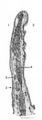
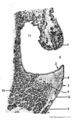
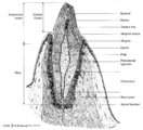
Scheme survey dorsal surface of tongue - human, adult | 1. foliate papillae (rudimentary in human); 2. circumvallate papillae; 3. foramen cecum; 4. palatine tonsils; 5. filiform papillae; 6. fungiform papillae; 7. epiglottis; 11. lingual tonsils | oral cavity | Poja Histology Collection - Oral Cavity Subset |
| 258 |
 |
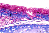
Scheme survey frontal section of larynx (human) | Stain: Azan. A pseudostratified epithelium covers the mucosa of the laryngeal cavity and vestibulum. At the vocal fold edge (3) the epithelium appears to be nonkeratinizing squamous. The connective tissue is rigidly attached to this edge and merges into the vocal ligament (dense elastic) and vocal m... | Vocal ligament; Laryngeal glands; Ventricular fold | Poja Histology Collection - Respiratory System Subset |
| 259 |
 |
Scheme survey lip - human, adult | 1. red (transitional) zone; 2. keratinized stratified squamous epithelium (outer side); 3. non-keratinized stratified squamous epithelium (inner side); 4. hair shaft; 5. hair follicle; 6. sebaceous gland; 7. cross-section of circumoral muscle; 8. labial glands with draining ducts; ... | oral cavity | Poja Histology Collection - Oral Cavity Subset |
| 260 |
 |
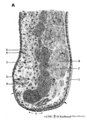
Scheme survey tooth (longitudinal section; human; canine) | Canine tooth in osseous alveolus. | oral cavity; pulp canal | Poja Histology Collection - Oral Cavity Subset |
| 261 |
 |
Scheme survey uvula - soft palate, human, adult | 1. nasal side - pseudostratified columnar ciliated epithelium (respiratory epithelium); 2. oral side - non-keratinized stratified squamous epithelium; 3. mixed glands (pharyngeal glands); 4. uvular muscle; 5. mucous palatine glands | oral cavity | Poja Histology Collection - Oral Cavity Subset |
| 262 |
 |
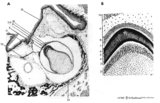
Secretory ameloblasts in tooth development - bell stage, gerbil, postnatal | Electronmicroscopy. Cross-sectioned distal sides of ameloblasts (secretion areas or Tomes' processes) with numerous organelles. The dark stained vesicles represent secretory granules contain among others amelogenin. At the right bottom corner intercellular spaces are filled with dark-stained organi... | oral cavity; enamel prisms | Poja Histology Collection - Oral Cavity Subset |
| 263 |
 |
Section of trachea (human, adult, high magnification) | Stain: Azan. On top: the epithelium (1) is pseudostratified with cilia and goblet cells (white droplets) on a distinct basement membrane (↓, undulating thick deep-red line) followed by a small proper lamina (2). The submucosa (3) starts where the fibrous tissue (elastic fibers reinforced with coll... | Pseudostratified epithelium ; Tracheal glands; Excretory duct; Perichondrium | Poja Histology Collection - Respiratory System Subset |
| 264 |
 |
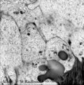
Small bronchus (golden hamster) | Electron microscopy. Two ciliated cells with nuclei (1). Supranuclearly electron-dense lysosomes (2, lipofucsin) are present, the apical parts contain mitochondria (3) and cross-sections of cilia (4) and microvilli (5) are visible in the lumen (*). Two mucoid goblet cells with packed secretion gran... | Ciliated epithelium | Poja Histology Collection - Respiratory System Subset |
| 265 |
 |
Small bronchus in the lung (human, adult) | Stain: Azan. Pseudostratified epithelium (1) with in between light-stained goblet cells and a blue stained basement membrane (↓) with the proper lamina. The thin smooth muscle layer (2) is purple stained and accompanies the mucosal layer. At (3) a small hyaline cartilage and seromucous bronchial g... | Small bronchus; Pseudostratified epithelium; Seromucous glands | Poja Histology Collection - Respiratory System Subset |
| 266 |
 |
Striated duct of nasal gland (rat) | Electron microscopy. At (1) lumen of duct, (2) indicates nucleus of lining epithelial cell. Note the abundancy and palisade-like arrangement of mitochondria (3) in the basolateral regions, in the light microscope visible as a fine striation, hence the name striated duct or intralobular duct. (4) ind... | Nasal vestibulum; Nasal glands; Striated duct; Intralobular duct; Seromucous glands | Poja Histology Collection - Respiratory System Subset |
| 267 |
 |
Sublingual gland (human) | Stain: Azan. The sublingual gland is a mixed gland and mucous cells are dominating (muco-serous acini). Intralobular ducts are present. Note one thick blue interlobular septum and few thinner intralobular ones. | oral cavity; seromucous gland | Poja Histology Collection - Oral Cavity Subset |
| 268 |
 |
Sublingual gland (human) | Stain: Azan. The sublingual gland is a mixed gland and characteristic in human is the presence of large areas of serous cells neighboring areas with mucous cells. Inter- and intra-lobular ducts are present. Note inter- and intra-lobular septa (blue) and scattered fat cells (white). | oral cavity; seromucous gland | Poja Histology Collection - Oral Cavity Subset |
| 269 |
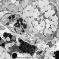 |
Submandibular gland (gerbil) | Electronmicroscopy. Upper two mucous cells of a seromucous acinus contain secretion droplets of different densities (light grey). Note partly fused mucous droplets. In the middle a serous cell cell with darkly stained granules. At the bottom a dense stained intercalated cell without granules. The ba... | oral cavity; seromucous gland | Poja Histology Collection - Oral Cavity Subset |
| 270 |
 |
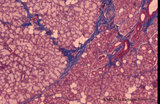
Submandibular gland (human) | Stain: Azan. The submandibular gland consists of mixed glands, i.e., the serous cells outnumber the mucous cells (seromucous acini). Locally crescent-shaped groups of serous cells (demi-lunes of Giannuzzi) are localized close to mucous cells to form an acinus.Intralobular ducts (left half) and thin ... | oral cavity; seromucous gland | Poja Histology Collection - Oral Cavity Subset |
| 271 |
 |
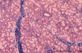
Submandibular gland (human) | Stain: Azan. Survey: on top capsule of dense connective tissue (blue) with septa dividing parenchyma of serous cells into lobules. At the bottom a large interlobular duct. Note white fat cells (adipocytes). | oral cavity; seromucous gland | Poja Histology Collection - Oral Cavity Subset |
| 272 |
 |
Submandibular gland (rat) | Electronmicroscopy. Darkly stained cells around the lumen of an intercalated duct and close to the lightly stained (partly degranulated) serous cells. | oral cavity; seromucous gland; intercalated duct | Poja Histology Collection - Oral Cavity Subset |
| 273 |
 |
Submucosa of trachea (human, adult) | Stain: Goldner trichrome. Mixed tracheal glands (1) of the submucosa (connective tissue light green) and bundles of smooth muscle fibers (2) close to the fibroelastic membrane between the cartilage edges. | Seromucous gland; Tracheal glands | Poja Histology Collection - Respiratory System Subset |
| 274 |
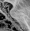 |
Surface of lung pleura (gerbil) | Scanning electron microscopy of visceral pleura. Alveoli (A) and capillaries (c) with scattered erythrocytes (artifactual due to preparation procedures). At (*) connective tissue and in between erythrocytes. The visceral surface is covered by a continuous sheet of flat squamous mesothelial cells (1,... | Visceral pleura; Mesothelium | Poja Histology Collection - Respiratory System Subset |
| 275 |
 |
Surface of olfactory epithelium (rat) | Electron microscopy. Bottom left shows an olfactory bulb (1, vesicle) with cross-sectioned basal bodies. Right side part of the apex of a supporting cell (2) with microvilli (3). Parallel to the surface of the epithelium one long olfactory cilium (4) and several other cross-sectioned ones are detect... | Olfactory epithelium; Olfactory vesicle | Poja Histology Collection - Respiratory System Subset |