John A. Moran Eye Center Neuro-Ophthalmology Collection: A variety of lectures, videos and images relating to topics in Neuro-Ophthalmology created by faculty at the Moran Eye Center, University of Utah, in Salt Lake City.
NOVEL: https://novel.utah.edu/
TO
Filters: Collection: "ehsl_novel_jmec"
| Identifier | Title | Description | Subject | ||
|---|---|---|---|---|---|
| 226 |
 |
ophthalmoscope | How to Use the Direct Ophthalmoscope in an Exam | Demonstration of using the direct ophthalmoscope to examine the optic disc. Covers hand placement , which eye to use, and distance from patient. | Direct Ophthalmoscope; Examination, Ocular |
| 227 |
 |
pulse | Spontaneous Venous Pulsations | This clips shows a spontaneous venous pulsation viewed during an ocular examination. | Spontaneous Venous Pulsations; Examination, Ocular; AVP Optic Nerve |
| 228 |
 |
sensory | Basic Neurologic Exam: Sensory | Demonstration of a sensory examination. | Neurology; Examinations |
| 229 |
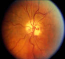 |
shunt vessel meningioma | Shunt Vessel Meningioma | RETINO-CHOROIDAL (OPTO-CILIARY) COLLATERAL VESSELS: (also known as Retinal-choroidal venous collaterals, opticociliary veins or ciliary shunt vessels) Retino-choroidal collaterals are potential telangiectatic connections between the retina and choroidal circulation. Although sometimes called "shunts... | Shunt Vessels (Meningioma) |
| 230 |
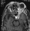 |
shunt vessel meningioma MRI | Shunt Vessel Meningioma - MRI | Meningiomas block venous egress and open potential venous channels known as retinochoroidal (optociliary) collateral vein. This meningioma extends from the back of the globe through the optic canal. | Shunt Vessels (Meningioma) |
| 231 |
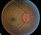 |
stage 2 | Stage 2 - Papilledema | Papilledema Stages; Raised Intracranial Pressure | |
| 232 |
 |
station_and_gait | Basic Neurologic Exam: Station and Gait | Demonstration of a station and gait examination. | Neurology; Examinations |
| 233 |
 |
trigeminal_nerve_exam | Trigeminal Nerve Exam | Explanation of a trigeminal nerve exam. | Trigeminal Nerve |
| 234 |
 |
visual_acuity | Measuring Visual Acuity | Demonstration on self of visual acuity exam, using a standard card. | Visual Acuity; Examination, Ocular |
| 235 |
 |
walsh_2000_c30 | The Wall-Eyed Potato Farmer | Young man presenting with apparent episodic neurologic evants that initially was thought to be multiple sclerosis, but as time went on, he had progressive changes in his neurologic exam and in his imaging findings. Brain biopsy revealed Gliomatosis cerebri. Anatomy: Brain Stem; Pons; Midbrain. Patho... | Gliomatosis Cerebri; Intracranial Tumors; Bilateral Internuclear Ophthalmoplegia |
| 236 |
 |
weinberg_1.pdf | Dysthyroid Optic Neuropathy: A Preventable Cause of Blindness | Dysthyroid Optic Neuropathy (DON) is a treatable cause of visual loss in ~5% of pts w/ ted. Monitor closely those pts with risk factors (proptosis, tight orbit, restricted motility, strabismus, smoker, diabetic). Oral prednisone is often effective, but frequent relapses after tapering. Orbital xrt ... | Dysthyroid Ophthalmopathy; Thyroid Orbitopathy; Thyroid Eye Disease; Thyroid Associated Ophthalmopathy (TAO); Graves' Disease; Restrictive Orbitopathy |