The Health Education Assets Library (HEAL) is a collection of over 22,000 freely available digital materials for health sciences education. The collection is now housed at the University of Utah J. Willard Marriott Digital Library.
TO
Filters: Collection: "ehsl_heal" Format: image
| Title | Description | Subject | Collection | ||
|---|---|---|---|---|---|
| 226 |
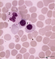 |
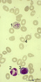
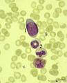
Orthochromatic erythroblasts in bone marrow smear (human) | Stain: May-Grnwald-Giemsa (MGG). Two orthochromatic erythroblasts (1) with condensed nuclear chromatin and a transition of cytoplasmic stain towards the color of normal erythrocytes. (2) a segmented neutrophilic granulocyte. | Poja Histology Collection - Blood & Bone Marrow Subset | |
| 227 |
 |
Orthochromatic erythroblast and lymphocyte in bone marrow smear (human) | Stain: May-Grnwald-Giemsa (MGG). The cytoplasm of this orthochromatic erythroblast (2) shows a faint polychromatic tint. The chromatin is arranged in clumps and stains deeply. (2) activated lymphocyte. | Poja Histology Collection - Blood & Bone Marrow Subset | |
| 228 |
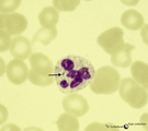 |
Normal mature neutrophilic granulocyte in peripheral blood smear (human) | Stain: May-Grnwald-Giemsa (MGG). Neutrophilic granulocyte with five nuclear lobes or segments, (not hypersegmented). The arrow indicates a drumstick. Note the fine dust-like granules. | Poja Histology Collection - Blood & Bone Marrow Subset | |
| 229 |
 |
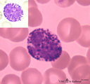
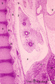
Mast cell | Mast cells (mastocytes) are oval (12 m) or spindle-shaped and are frequently found perivascularly or perineurally. The cytoplasm is provided with a moderate amount of organelles and small thin or blunt microvilli at the surface. Most obvious is the presence of large granules varying in shape and siz... | Poja Histology Collection - Blood & Bone Marrow Subset | |
| 230 |
 |
Toxic granulation and vacuolization in neutrophilic myelocytes in peripheral blood smear (human) | Stain: May-Grnwald-Giemsa (MGG). The myelocyte (1) and the metamyelocyte (2) show toxic granulation, vacuolisation and cytoplasmic swelling. Toxic granulation is characterized by violet-purple granules of varying size in the cytoplasm; generally admixed with normal pink granules. The phenomenon occu... | Poja Histology Collection - Blood & Bone Marrow Subset | |
| 231 |
 |
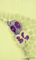
Howell-Jolly bodies in reticulocytes in peripheral blood smear (human) | Stain: May-Grnwald-Giemsa (MGG). The Howell-Jolly-bodies (1) in the reticulocyte are remnants of the nucleus (nuclear chromatin granules) of an erythroblast during anomalous development due to vitamin B12 deficiency, folic acid deficiency or in megaloblastic and hemolytic anemias. (2) Neutrophil wit... | Poja Histology Collection - Blood & Bone Marrow Subset | |
| 232 |
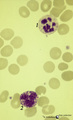 |
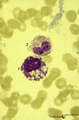
Neutrophilic and basophilic granulocytes in peripheral blood smear (human) | Stain: May-Grnwald-Giemsa (MGG). (1) A hypersegmented (>5 segments) neutrophilic granulocyte with clear fine granules. (2) Represents a mature basophilic granulocyte with clear distinguishable, coarse purple granules and a few vacuoles (because the granules dissolve in water during the staining proc... | Poja Histology Collection - Blood & Bone Marrow Subset | |
| 233 |
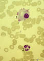 |
Flaming plasma cell in peripheral blood smear (human) | Stain: May-Grnwald-Giemsa (MGG). The so-called flaming plasma cell (1) is characterized by fiery fringes, which are formed by pseudopodic cytoplasmic projections (arrows) that stain with carmin red. These peripheral cytoplasmic spots contain numerous dilated endoplasmic reticulum cisterns, which are... | Poja Histology Collection - Blood & Bone Marrow Subset | |
| 234 |
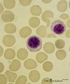 |
Plasmacytoid lymphocytes (activated B cells) in peripheral blood smear (human) | Stain: May-Grnwald-Giemsa (MGG). Two different plasmacytoid lymphocytes or activated young B cells (up to 15 μm) contain a dark-stained nucleus and a slightly basophilic cytoplasm with a kind of a 'nuclear hof' indicating the Golgi area. These cells will develop into plasma cells. | Poja Histology Collection - Blood & Bone Marrow Subset | |
| 235 |
 |
Monoblast in bone marrow smear (human) | Stain: May-Grnwald-Giemsa (MGG). The monoblast (1) has a large nucleus with an irregular border, nucleoli and fine disperse chromatin, and a basophilic cytoplasm with no or a limited number of granules. (2) Two neutrophilic band forms. | Poja Histology Collection - Blood & Bone Marrow Subset | |
| 236 |
 |
Basophilic granulocyte in peripheral blood smear (human) | Stain: May-Grnwald-Giemsa (MGG). The basophilic granulocyte is characterized by large, coarse, aggregated dark purple granules. The nuclear lobes are usually not very well visible and masked by the granules (see also inset). | Poja Histology Collection - Blood & Bone Marrow Subset | |
| 237 |
 |
Dividing cells (mitosis) in bone marrow smear (human) | Stain: May-Grnwald-Giemsa (MGG). (1) Shows a dividing cell (mitotic figure) possibly an erythroblast cell type. (2) Shows two segmented neutrophils. | Poja Histology Collection - Blood & Bone Marrow Subset | |
| 238 |
 |
Plasma cell | Scheme electron microscopy. The mature plasma cell or plasmacyte (12-15 m) shows an eccentric nucleus (1) with a characteristic chunky distribution of heterochromatin along the inner nuclear membrane ('spoke-wheel' effect in light microscopy). Juxta-nuclearly an elaborate Golgi area (2) with secreti... | Poja Histology Collection - Blood & Bone Marrow Subset | |
| 239 |
 |
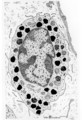
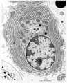
Basophilic granulocyte | Scheme electron microscopy. Basophils are non-phagocytic granulocytes that account for 0.5% to 1.0% of the circulating white blood cells. Their granulated cytoplasm stains with basic dyes, hence the name basophil. Electron microscopy reveals a multilobed nucleus (1); few mitochondria (2); numerous ... | Poja Histology Collection - Blood & Bone Marrow Subset | |
| 240 |
 |
Eosinophilic myelocyte and neutrophilic myelocyte in bone marrow smear (human) | Stain: May-Grnwald-Giemsa (MGG). The eosinophilic myelocyte (1) contains brown-like granules (not orange) in the bluish basophilic cytoplasm. Nucleoli are still visible. Maturation of eosinophils parallels that of neutrophils except for the production of the secondary, specific granules in myelocyte... | Poja Histology Collection - Blood & Bone Marrow Subset | |
| 241 |
 |
Mature erythrocytes (spleen, gerbil) | Scanning electron microscopy. Normal mature erythrocytes (1) are mostly biconcave and discoid. They might change their forms due to mechanical pressures. (2) a mature lymphocyte recognizable by many short stubby microvilli at the surface. (3) granulocytes are larger with fewer but longer microvilli.... | Poja Histology Collection - Blood & Bone Marrow Subset | |
| 242 |
 |
Olfactory epithelium in the nasal cavity (mammals) | Three-dimensional scheme electron microscopy. Six olfactory cells (1) surround one massive central supporting cell (2). A third, basal cell (3) is located at the basal plate aposing to the supporting cell. These olfactory cells are tall and slender bipolar nerve cells with chemoreceptive functions. ... | Pseudostratified epithelium; Olfactory epithelium; Basal cells; Olfactory vesicle; Bipolar neuron | Poja Histology Collection - Respiratory System Subset |
| 243 |
 |
Keratin 7 staining in dysplastic epithelium of a bronchus (human, adult) | Stain: anti-keratin 7 antibody (Pan-Ck 7) immunoperoxidase staining (with aminoethylcarbazole (AEC) substrate). A red-brown staining with AEC indicates a positive reaction for cytokeratin 7. Disturbances in the growth and maturing might result in dysplasia of the epithelium with a changed reaction p... | Immunoperoxidase; Immuno-reaction | Poja Histology Collection - Respiratory System Subset |
| 244 |
 |
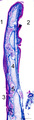
Pseudoglandular period of developing lung (human, embryo, low magnification) | Stain: Hematoxylin and eosin. Cross-sectioned future bronchial tubes (1), the surrounding mesenchyme becomes more condensed around the epithelium. The mesoderm of the future visceral pleura (2) as well as the future parietal pleura (3) and (4) indicates pleural cavity. The cartilagineous spinal colu... | Lung development; Visceral pleura; Parietal pleura; Pseudoglandular period; Bronchial tubes; Mesenchyme | Poja Histology Collection - Respiratory System Subset |
| 245 |
 |
Pseudoglandular period of developing lung (human, embryo, low magnification) | Stain: Trichrome (Goldner). Cross-sectioned future bronchial tubes (1) of varying sizes. Note the apical position of the nuclei in these epithelial cells with light-stained basal parts (glycogen). The surrounding mesenchyme becomes more condensed (↓) around the epithelium and in between numerous ... | Lung development; Visceral pleura; Pseudoglandular period; Bronchial tubes; Mesenchyme | Poja Histology Collection - Respiratory System Subset |
| 246 |
 |
Nasal glands of the nasal vestibulum (rat) | Electron microscopy. At the top part of a striated draining duct with a wide lumen (*); these cuboidal ductal cells (1) contain many basolaterally located mitochondria. The ductal cells are enforced by interstitial fibroblast (2) and a capillary (4). Neighboring serous gland cells (5) contain dark s... | Nasal vestibulum; Seromucous glands; Nasal glands; Serous cells; Secretion granules; Intercalated duct; Striated duct | Poja Histology Collection - Respiratory System Subset |
| 247 |
 |
Small bronchus in the lung (human, adult) | Stain: Azan. Pseudostratified epithelium (1) with in between light-stained goblet cells and a blue stained basement membrane (↓) with the proper lamina. The thin smooth muscle layer (2) is purple stained and accompanies the mucosal layer. At (3) a small hyaline cartilage and seromucous bronchial g... | Small bronchus; Pseudostratified epithelium; Seromucous glands | Poja Histology Collection - Respiratory System Subset |
| 248 |
 |
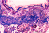
Survey of epiglottis (human) | Stain: Azan. Centrally light-stained elastic cartilage (4). Lingual side (2) is covered with squamous epithelium. At the laryngeal side (1) the squamous epithelium usually turns into respiratory epithelium with seromucous laryngeal glands (5). Note also lymphoid aggregations in this area (3 →). | Oral cavity; Laryngeal glands | Poja Histology Collection - Respiratory System Subset |
| 249 |
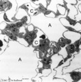 |
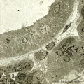
The terminal part of the airway: alveoli (dog) | Electron microscopy (low magnification). (A) indicate alveoli; (C) indicate capillaries. (1) type I alveolar cell (pneumocyte I, squamous alveolar cell); (2) type II alveolar cell (pneumocyte II, great alveolar cell). (3) alveolar macrophage. (4) endothelium of capillary. | Pneumocyte I; Pneumocyte II; Alveolar macrophage | Poja Histology Collection - Respiratory System Subset |
| 250 |
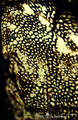 |
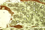
Capillary system of a lung alveolus (cat) | Stain: specimen injected with India ink via the pulmonary system. In an en face view of the alveolar wall the black- and granular-stained capillary plexus is well shown. The larger vessels represent branches of the pulmonary arteriole. | Poja Histology Collection - Respiratory System Subset |