The Health Education Assets Library (HEAL) is a collection of over 22,000 freely available digital materials for health sciences education. The collection is now housed at the University of Utah J. Willard Marriott Digital Library.
TO
Filters: Collection: "ehsl_heal"
| Title | Description | Subject | Collection | ||
|---|---|---|---|---|---|
| 226 |
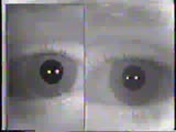 |
Cranial Nerve Exam: Abnormal Examples: Cranial Nerves 2 & 3 - Pupillary Light Reflex | The swinging flashlight test is used to show a relative afferent pupillary defect or a Marcus Gunn pupil of the left eye. The left eye has perceived less light stimulus (a defect in the sensory or afferent pathway) then the opposite eye so the pupil dilates with the same light stimulus that caused c... | Cranial Nerve Examination; Marcus Gunn Pupil | NeuroLogic Exam: An Anatomical Approach |
| 227 |
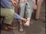 |
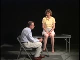
Coordination Exam: Abnormal Examples: Heel-to-shin (includes Spanish audio & captions) | The patient with ataxia of the lower extremity will have difficulty placing the heel on the knee with a side-to-side irregular over- and undershooting as the heel is advanced down the shin. Dysmetria on heel-to-shin can be seen in midline ataxia syndromes as well as cerebellar hemisphere disease so ... | Coordination Examination; Heel-shin Test | NeuroLogic Exam: An Anatomical Approach |
| 228 |
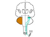 |
Coordination Exam: Anatomy: Spinocerebellum (x2) (includes Spanish audio & captions) | The 2nd subdivision of the cerebellum is the spinocerebellum. This system consists of the connections between the cutaneous and proprioceptive information coming from the spinal cord to the vermis and paravermis regions with corrective feedback predominantly to the muscles of truncal stability and g... | Coordination Examination; Anterior Lobe of Cerebellum; Spinocerebellum | NeuroLogic Exam: An Anatomical Approach |
| 229 |
|
Coordination Exam: Anatomy: Spinocerebellum (includes Spanish audio & captions) | The 2nd subdivision of the cerebellum is the spinocerebellum. This system consists of the connections between the cutaneous and proprioceptive information coming from the spinal cord to the vermis and paravermis regions with corrective feedback predominantly to the muscles of truncal stability and g... | Coordination Examination; Anterior Lobe of Cerebellum; Spinocerebellum | NeuroLogic Exam: An Anatomical Approach |
| 230 |
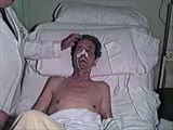 |
Cranial Nerve Exam: Abnormal Examples: Cranial Nerve 7 - Motor (x2) | The first patient has weakness of all the muscles of facial expression on the right side of the face indicating a lesion of the facial nucleus or the peripheral 7th nerve. The second patient has weakness of the lower half of his left face including the orbicularis oculi muscle but sparing the forehe... | NeuroLogic Exam: An Anatomical Approach | |
| 231 |
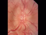 |
Cranial Nerve Exam: Abnormal Examples: Cranial Nerve 2 - Fundoscopy (x2) | The first photograph is of a fundus showing papilledema. The findings of papilledema include 1. Loss of venous pulsations 2. Swelling of the optic nerve head so there is loss of the disc margin 3. Venous engorgement 4. Disc hyperemia 5. Loss of the physiologic cup and 6. Flame shaped hemorrhages. Th... | Cranial Nerve Examination | NeuroLogic Exam: An Anatomical Approach |
| 232 |
 |
Cranial Nerve Exam: Normal Exam: Vergence | Vergence eye movements occur when the eyes move simultaneously inward (convergence) or outward (divergence) in order to maintain the image on the fovea that is close up or far away. Most often convergence is tested as part of the near triad. When a patient is asked to follow an object that is brough... | Cranial Nerve Examination; Vergence | NeuroLogic Exam: An Anatomical Approach |
| 233 |
|
Cranial Nerve Exam: Abnormal Examples: Cranial Nerve 7 - Motor | The first patient has weakness of all the muscles of facial expression on the right side of the face indicating a lesion of the facial nucleus or the peripheral 7th nerve. The second patient has weakness of the lower half of his left face including the orbicularis oculi muscle but sparing the forehe... | NeuroLogic Exam: An Anatomical Approach | |
| 234 |
 |
Aortocaval Fistula (Labeled) | Fistula between aorta and inferior vena cava. | Aortocaval Fistula | Royal College of Surgeons in Ireland Illustrations |
| 235 |
|
Resection and Primary Anastomosis of Sigmoid Colon (Labeled) | Colon resection. Primary anastomosis. | Primary Anastomosis; Resection | Royal College of Surgeons in Ireland Illustrations |
| 236 |
|
Mastectomy - Right Breast | Breast cancer. Incision site. Mastectomy. | Incision Site; Breast Cancer | Royal College of Surgeons in Ireland Illustrations |
| 237 |
|
Coordination Exam: Abnormal Examples: Heel-to-shin (x2) (includes Spanish audio & captions) | The patient with ataxia of the lower extremity will have difficulty placing the heel on the knee with a side-to-side irregular over- and undershooting as the heel is advanced down the shin. Dysmetria on heel-to-shin can be seen in midline ataxia syndromes as well as cerebellar hemisphere disease so ... | Coordination Examination; Heel-shin Test | NeuroLogic Exam: An Anatomical Approach |
| 238 |
 |
Coordination Exam: Abnormal Examples: Finger-to-nose (x2) (includes Spanish audio & captions) | Under (hypometria) and over (hypermetria) shooting of a target (dysmetria) and the decomposition of movement (the breakdown of the movement into its parts with impaired timing and integration of muscle activity) are seen with appendicular ataxia. NeuroLogic Exam has been supported by a grant from th... | Coordination Examination; Finger-to-nose Test | NeuroLogic Exam: An Anatomical Approach |
| 239 |
 |
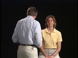
Coordination Exam: Normal Exam: Finger-to-nose (includes Spanish audio & captions) | The patient moves her pointer finger from her nose to the examiner's finger as the examiner moves his finger to new positions and tests accuracy at the furthest outreach of the arm. NeuroLogic Exam has been supported by a grant from the Slice of Life Development Fund at the University of Utah, th... | Coordination Examination; Finger-to-nose Test | NeuroLogic Exam: An Anatomical Approach |
| 240 |
 |
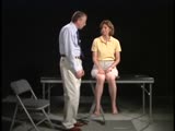
Coordination Exam: Normal Exam: Heel-to-shin (includes Spanish audio & captions) | The patient places her heel on the opposite knee then runs the heel down the shin to the ankle and back to the knee in a smooth coordinated fashion. NeuroLogic Exam has been supported by a grant from the Slice of Life Development Fund at the University of Utah, the Department of Pediatrics and the O... | Coordination Examination; Heel-shin Test | NeuroLogic Exam: An Anatomical Approach |
| 241 |
|
Resection and End Colostomy | Colon resection. End colostomy. Harmann's procedure. | End Colostomy; Colon Resection; Hartmann's Procedure | Royal College of Surgeons in Ireland Illustrations |
| 242 |
|
Needle stick | Picture of a needle stick in a plastic arm | Venipuncture | HEAL Reviewed Collection |
| 243 |
|
Equipment for venipuncture | This is an image of equipment for venipuncture. | Venipuncture | HEAL Reviewed Collection |
| 244 |
|
Gastrointestinal Tract (Labeled) | Gastrointestinal tract showing upper GI, ligament of Treitz, and lower GI. | Upper GI; Upper Gastrointestinal Tract; Ligament of Treitz; Lower GI; Lower Gastrointestinal Tract | Royal College of Surgeons in Ireland Illustrations |
| 245 |
|
Resection and Primary Anastomosis of Sigmoid Colon | Make private -- psd file. Colon resection. Primary anastomosis. | Primary Anastomosis; Resection | Royal College of Surgeons in Ireland Illustrations |
| 246 |
|
Male Urogenital Diaphragm | Urogenital diaphragm. Male. | Ischiopubic Ramus; Perineal Membrane; Urogenital Diaphragm; Perineal Body; Penile Veins; Prostatic Venous Plexus | Royal College of Surgeons in Ireland Illustrations |
| 247 |
|
Exposed Facial Nerve with Retracted Parotid Gland and Stylohyoid Muscle (Labeled) | Exposed facial nerve with retracted parotid gland and stylohyoid muscle. | Stylohyoid Muscle | Royal College of Surgeons in Ireland Illustrations |
| 248 |
 |
Eye - Rods and Cones | Rod and cone photoreceptors have their cell bodies in the outer nuclear layer. Between these cell bodies and the retinal pigment epithelium are the photoreceptor inner segments (where proteins are synthesized) and the outer segments (the light sensitive portion). The inner segments of cones are cone... | UCLA Histology | |
| 249 |
 |
Uterus | This relatively brief phase of the endometrial cycle consists of shrinkage of the endometrium and intraendometrial hemorrhage and fragmentation. The myometrium is located to the left. A higher magnification of the ischaemic endometrium is shown in image 226-10-1. UCLA Histology Collection. | ischaemic endometrium; Uterus | UCLA Histology |
| 250 |
 |
Kidney | In this section through the renal papilla, identify the terminal collecting tubules (lined by columnar epithelium). Also note a straight segment of the loop of Henle, many thin loops of Henle, and cross-sections of the vasa recta. UCLA Histology Collection. | Kidney; renal papilla; terminal collecting tubules | UCLA Histology |