John A. Moran Eye Center Neuro-Ophthalmology Collection: A variety of lectures, videos and images relating to topics in Neuro-Ophthalmology created by faculty at the Moran Eye Center, University of Utah, in Salt Lake City.
NOVEL: https://novel.utah.edu/
TO
Filters: Type: "Image" Collection: ehsl_novel_jmec
1 - 25 of 3
| Identifier | Title | Description | Subject | ||
|---|---|---|---|---|---|
| 1 |
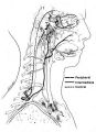 |
Figure-04 | Anatomy of the Oculosympathetic Pathway | Anatomy of the oculosympathetic pathway. (Maloney WF, Younge BR, Moyer NJ: Evaluation of the causes and accuracy of pharmacologic localization in Horner's syndrome. Am J Ophthalmol 1980;90:394-402, Ophthalmic Publishing Company with permission.) | Anatomy of the Oculosympathetic Pathway; Horner's Syndrome |
| 2 |
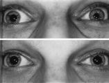 |
Figure-21 | Left-sided Dilation Lag in a Man with Horner's Syndrome | Left-sided dilation lag in a 29-year-old man with Horner's syndrome caused by a posterior mediastinal ganglioneuroma. Note that the degree of anisocoria is greater after 5 seconds in darkness (top) compared with findings after 15 seconds in darkness (bottom). | Diagnosis, Horner Syndrome; Physiopathology, Horner Syndrome; Reflex, Pupillary; Dilation Lag; Horner's Syndrome |
| 3 |
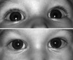 |
Figure-22 | Right-sided Pseudo-Horner's Syndrome | Right-sided pseudo-Horner's syndrome in an 8-month-old infant referred because her mother had noted a larger pupil on the left for a few months and her pediatrician thought the right upper lid was droopy. Both pupils reacted normally to light and darkness, the degree of anisocoria was similar in bot... | Infant; Diagnosis, Horner Syndrome; Etiology, Horner Syndrome; Horner's Syndrome |
1 - 25 of 3