TO
Filters: Type: "Image" Collection: ehsl_novel_*
| Title | Curriculum | Description | Subject | Collection | ||
|---|---|---|---|---|---|---|
| 1 |
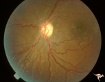 |
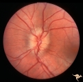
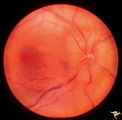
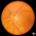
ID05a Post Papilledema Optic Atrophy from Pseudotumor Cerebri | Left eye, October 1999, Post papilledema optic atrophy from pseudotumor cerebri. Note optociliary veins in both discs. Gliosis and partial pallor following long standing papilledema and intracranial pressure. Anatomy: Optic disc. Pathology: Post papilledema atrophy and gliosis from long standing el... | Optic Disc Atrophy with Special Features; Atrophy with Arteriolar Sheathing; Gliosis and Pseudodrusen; Post Papilledema Atrophy | Neuro-Ophthalmology Virtual Education Library: William F. Hoyt Neuro-Ophthalmology Collection: https://novel.utah.edu/Hoyt/ | |
| 2 |
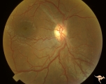 |
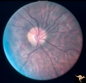
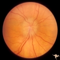
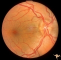
ID05b Post Papilledema Optic Atrophy from Pseudotumor Cerebri | Right eye, October 1999, Post papilledema optic atrophy from pseudotumor cerebri. Note optociliary veins in both discs. Gliosis and partial pallor following long standing papilledema and intracranial pressure. Anatomy: Optic disc. Pathology: Post papilledema atrophy and gliosis from long standing el... | Optic Disc Atrophy with Special Features; Atrophy with Arteriolar Sheathing; Gliosis and Pseudodrusen; Post Papilledema Atrophy | Neuro-Ophthalmology Virtual Education Library: William F. Hoyt Neuro-Ophthalmology Collection: https://novel.utah.edu/Hoyt/ | |
| 3 |
 |
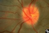
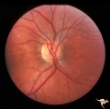
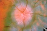
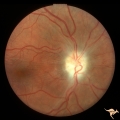
Isolated Optic Neuritis/Neuropathy | Papilledema in pseudotumor cerebri may result in adjacent choroidal or retinal folds. | Pseudotumor Cerebri/Papilledema; Edema | Neuro-Ophthalmology Virtual Education Library: NOVEL http://NOVEL.utah.edu | |
| 4 |
 |
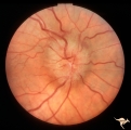
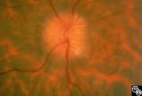
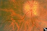
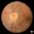
Isolated Optic Neuritis/Neuropathy | This image demonstrates Paton's lines in a 34-year-old patient with pseudotumor cerebri and chronic papilledema. | Pseudotumor Cerebri/Papilledema; Edema | Neuro-Ophthalmology Virtual Education Library: NOVEL http://NOVEL.utah.edu | |
| 5 |
 |
Unilateral Papilledema | Right eye. Has papilledema. Patient has pseudotumor cerebri. 24 year old obese woman. Anatomy: Optic disc. Pathology: Unilateral papilledema. Disease/Diagnosis: Idiopathic intracranial hypertension, pseudotumor cerebri. Clinical: Woman, headache, transient visual obscurations. | Unilateral Papilledema | Neuro-Ophthalmology Virtual Education Library: William F. Hoyt Neuro-Ophthalmology Collection: https://novel.utah.edu/Hoyt/ | |
| 6 |
 |
Unilateral Papilledema | Left eye. Has papilledema. Patient has Pseudotumor cerebri. Woman. Anatomy: Optic disc. Pathology: Unilateral papilledema. Disease/Diagnosis: Idiopathic intracranial hypertension, pseudotumor cerebri. Clinical: Woman, headache, transient visual obscuration. | Unilateral Papilledema | Neuro-Ophthalmology Virtual Education Library: William F. Hoyt Neuro-Ophthalmology Collection: https://novel.utah.edu/Hoyt/ | |
| 7 |
 |
Unilateral Papilledema | Unilateral papilledema in pseudotumor cerebri. Right eye. Has no cup. 27 year old white woman. Anatomy: Optic disc. Pathology: Unilateral papilledema. Disease/Diagnosis: Idiopathic intracranial hypertiension, pseudotumor cerebri. Clinical: Woman, headache. | Unilateral Papilledema | Neuro-Ophthalmology Virtual Education Library: William F. Hoyt Neuro-Ophthalmology Collection: https://novel.utah.edu/Hoyt/ | |
| 8 |
 |
Unilateral Papilledema | Right eye. Has no optic cup. Patient has pseudotumor cerebri. Woman. Anatomy: Optic disc. Pathology: Unilateral papilledema. Disease/Diagnosis: Idiopathic intracranial hypertension, pseudotumor cerebri. Clinical: Woman, headache, transient visual obscurations. | Unilateral Papilledema | Neuro-Ophthalmology Virtual Education Library: William F. Hoyt Neuro-Ophthalmology Collection: https://novel.utah.edu/Hoyt/ | |
| 9 |
 |
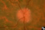
Isolated Optic Neuritis/Neuropathy | This 42-year-old male with pseudotumor cerebri and chronic papilledema demonstrated refractile bodies, which can be seen with chronic optic disc edema. This image exhibits decreased disc edema and resolution of the refractile bodies OD after therapy. Pair with 96_01, 96_02, 96_03, 96_05, and 96_06. | Pseudotumor Cerebri/Papilledema; Edema | Neuro-Ophthalmology Virtual Education Library: NOVEL http://NOVEL.utah.edu | |
| 10 |
 |
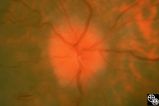
Isolated Optic Neuritis/Neuropathy | This 42-year-old male with pseudotumor cerebri and chronic papilledema demonstrated refractile bodies, which can been seen with chronic optic disc edema. This image demonstrates later recurrence of the refractile bodies with worsening papilledema OD. Pair with 96_01, 96_02, 96_03, 96_04, and 96_06. | Pseudotumor Cerebri/Papilledema; Edema | Neuro-Ophthalmology Virtual Education Library: NOVEL http://NOVEL.utah.edu | |
| 11 |
 |
Isolated Optic Neuritis/Neuropathy | This 42-year-old male with pseudotumor cerebri and chronic papilledema demonstrated refractile bodies, which can be seen with chronic optic disc edema. This image demonstrates later recurrence of the refractile bodies with worsening papilledema OD. Pair with 96_01, 96_02, 96_03, 96_04, and 96_05. | Pseudotumor Cerebri/Papilledema; Edema | Neuro-Ophthalmology Virtual Education Library: NOVEL http://NOVEL.utah.edu | |
| 12 |
 |
Isolated Optic Neuritis/Neuropathy | This 42-year-old male with pseudotumor cerebri and chronic papilledema demonstrated refractile bodies, which can been seen with chronic optic disc edema. This image exhibits decreased disc edema and resolution of the refractile bodies OD after therapy. Pair with 96_01, 96_02, 96_04, 96_05, and 96_06... | Pseudotumor Cerebri/Papilledema; Edema | Neuro-Ophthalmology Virtual Education Library: NOVEL http://NOVEL.utah.edu | |
| 13 |
 |
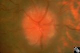
Papilledema and Resolution (PowerPoint) | curriculum_fellow; KBDpapilledemaandresolution; IC-C6bi-optic-disc-edema; KBDpapilledema | This 42-year-old male with pseudotumor cerebri and chronic papilledema demonstrated refractile bodies, which can be seen with chronic optic disc edema. This image shows the chronic papilledema at presentation, with associated refractile hyaline bodies at the disc periphery in both eyes. | Pseudotumor Cerebri/Papilledema; Edema; Papilledema and Resolution | Neuro-ophthalmology Virtual Education Library: NOVEL http://NOVEL.utah.edu |
| 14 |
 |
Unilateral papilledema | Unilateral papilledema in Pseudotumor cerebri in patient with elevated intracranial pressure. Right eye. Anatomy: Optic disc. Pathology: Unilateral papilledema. Disease/Diagnosis: Idiopathic intracranial hypertension (pseudotumor cerebri). Clinical: Transient monocular blindness (transient visual ob... | Unilateral Papilledema | Neuro-Ophthalmology Virtual Education Library: William F. Hoyt Neuro-Ophthalmology Collection: https://novel.utah.edu/Hoyt/ | |
| 15 |
 |
Unilateral Papilledema | Unilateral papilledema in Pseudotumor cerebri in patient with elevated intracranial pressure. Right eye. Anatomy: Optic disc. Pathology: Unilateral papilledema. Disease/Diagnosis: Idiopathic intracranial hypertension (pseudotumor cerebri). Clinical: Transient monocular blindness (transient visual ob... | Unilateral Papilledema | Neuro-Ophthalmology Virtual Education Library: William F. Hoyt Neuro-Ophthalmology Collection: https://novel.utah.edu/Hoyt/ | |
| 16 |
 |
Isolated Optic Neuritis/Neuropathy | This 42-year-old male with pseudotumor cerebri and chronic papilledema demonstrated refractile bodies, which can be seen with chronic optic disc edema. This image shows the chronic papilledema at presentation, with associated refractile hyaline bodies at the disc periphery in both eyes. Pair with 96... | Pseudotumor Cerebri/Papilledema; Edema | Neuro-Ophthalmology Virtual Education Library: NOVEL http://NOVEL.utah.edu | |
| 17 |
 |
Isolated Optic Neuritis/Neuropathy | This 42-year-old male with pseudotumor cerebri and chronic papilledema demonstrated refractile bodies, which can be seen with chronic optic disc edema. This image shows the chronic papilledema at presentation, with associated refractile hyaline bodies at the disc periphery in both eyes. Pair with 96... | Pseudotumor Cerebri/Papilledema; Edema | Neuro-Ophthalmology Virtual Education Library: NOVEL http://NOVEL.utah.edu | |
| 18 |
 |
Unilateral Papilledema | Unilateral papilledema in Pseudotumor cerebri in patient with elevated intracranial pressure. Left eye. Has no optic cup. Disc is flat. Anatomy: Optic disc. Pathology: Unilateral papilledema. Disease/Diagnosis: Idiopathic intracranial hypertension (pseudotumor cerebri). Clinical: Transient monocular... | Unilateral Papilledema | Neuro-Ophthalmology Virtual Education Library: William F. Hoyt Neuro-Ophthalmology Collection: https://novel.utah.edu/Hoyt/ | |
| 19 |
 |
Unilateral Papilledema | curriculum_fellow; KBDpapilledema; KBDunilateralpapilledema; KBDpseudotumorcerebri; IC-E10ai3a-idiopathic-intracranial-hypertension; IC-D1i-papilladema-secondary-to-raised-intracranial-pressure | Unilateral papilledema in Pseudotumor cerebri. Right eye. Has no cup. Woman. Anatomy: Optic disc. Pathology: Unilateral papilledema. Disease/Diagnosis: Idiopathic intracranial hypertension, pseudotumor cerebri. Clinical: Woman, headache. | Unilateral Papilledema; Congenital Blurred Disc | Neuro-Ophthalmology Virtual Education Library: William F. Hoyt Neuro-Ophthalmology Collection: https://novel.utah.edu/Hoyt/ |
| 20 |
 |
Bilateral Papilledema with Pseudotumor Cerebri | curriculum_fellow; KBDpapilledema; KBDpseudotumorcerebri; IC-E10ai3a-idiopathic-intracranial-hypertension; IC-D1i-papilladema-secondary-to-raised-intracranial-pressure | Right eye. Mild bilateral papilledema in a 7 year old boy. Cause of swelling unknown. Growth failure treated with thyroid medication. Anatomy: Optic disc. Pathology: Bilateral papilledema. Disease/Diagnosis: Intracranial hypertension due to treatment of growth failure with thyroid medicaltion. Clini... | Bilateral Papilledema; Pseudotumor Cerebri; Idiopathic Intracranial Hypertension | Neuro-Ophthalmology Virtual Education Library: William F. Hoyt Neuro-Ophthalmology Collection: https://novel.utah.edu/Hoyt/ |
| 21 |
 |
Bilateral Papilledema with Pseudotumor Cerebri | curriculum_fellow; KBDpapilledema; KBDpseudotumorcerebri; IC-E10ai3a-idiopathic-intracranial-hypertension; IC-D1i-papilladema-secondary-to-raised-intracranial-pressure | Left eye. Mild bilateral papilledema in a 7 year old boy. Cause of swelling unknown. Growth failure treated with thyroid medication. Anatomy: Optic disc. Pathology: Bilateral papilledema. Disease/Diagnosis: Intracranial hypertension due to treatment of growth failure with thyroid medication. Clinica... | Bilateral Papilledema; Pseudotumor Cerebri; Idiopathic Intracranial Hypertension | Neuro-Ophthalmology Virtual Education Library: William F. Hoyt Neuro-Ophthalmology Collection: https://novel.utah.edu/Hoyt/ |
| 22 |
 |
Bilateral Papilledema with Pseudotumor Cerebri | curriculum_fellow; KBDpapilledema; KBDpseudotumorcerebri; IC-E10ai-intracranial-hypertension; IC-D1i-papilladema-secondary-to-raised-intracranial-pressure | Chronic appearance of swelling in right eye. 29 year old woman. Bilateral papilledema. Anatomy: Optic disc. Pathology: Bilateral papilledema. Disease/Diagnosis: Intracranial hypertension due to treatment of growth failure with thyroid medication. Clinical: symptoms: headache, signs: bilateral papill... | Bilateral Papilledema; Pseudotumor Cerebri; Raised Intracranial Pressure | Neuro-Ophthalmology Virtual Education Library: William F. Hoyt Neuro-Ophthalmology Collection: https://novel.utah.edu/Hoyt/ |
| 23 |
 |
ID03a Post Papilledema Atrophy with Marked Gliosis | Post papilledema atrophy with marked gliosis in a patient with pseudotumor cerebri. Patient weighed over 300 pounds. Right eye blind. 1981. Right eye. Pair with ID_3b. Anatomy: Optic disc. Pathology: Post papilledema atrophy and gliosis from long standing elevated intracranial pressure. Disease/ Dia... | Optic Disc Atrophy with Special Features; Atrophy with Arteriolar Sheathing; Gliosis and Pseudodrusen; Post Papilledema Atrophy | Neuro-Ophthalmology Virtual Education Library: William F. Hoyt Neuro-Ophthalmology Collection: https://novel.utah.edu/Hoyt/ | |
| 24 |
 |
ID03b Post Papilledema Atrophy with Marked Gliosis | Post papilledema atrophy with marked gliosis in a patient with pseudotumor cerebri. Patient weighed over 300 pounds. Left eye has visual field defects. 1981, right eye, pair with ID_3a. Anatomy: Optic disc. Pathology: Post papilledema atrophy and gliosis from long standing elevated intracranial pres... | Optic Disc Atrophy with Special Features; Atrophy with Arteriolar Sheathing; Gliosis and Pseudodrusen; Post Papilledema Atrophy | Neuro-Ophthalmology Virtual Education Library: William F. Hoyt Neuro-Ophthalmology Collection: https://novel.utah.edu/Hoyt/ | |
| 25 |
 |
ID04a Post Papilledema Atrophy with Marked Gliosis | Post papilledema atrophy with marked gliosis in a patient with pseudotumor cerebri, 1985, right eye, pair with ID_4b, Note "high water" marks in peripapillary pigment epithelial layer. Anatomy: Optic disc. Pathology: Post papilledema atrophy and gliosis from long standing elevated intracranial press... | Optic Disc Atrophy with Special Features; Atrophy with Arteriolar Sheathing; Gliosis and Pseudodrusen; Post Papilledema Atrophy | Neuro-Ophthalmology Virtual Education Library: William F. Hoyt Neuro-Ophthalmology Collection: https://novel.utah.edu/Hoyt/ |