|
|
Title | Date | Type | Setname |
| 101 |
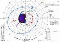 |
Chiasmal Compression - Vascular - KineticOS | 2024-07 | Image | ehsl_novel_nrm |
| 102 |
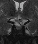 |
Chiasmal Compression - Vascular - l | 2024-07 | Image | ehsl_novel_nrm |
| 103 |
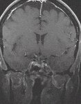 |
Chiasmal Compression - Vascular - m | 2024-07 | Image | ehsl_novel_nrm |
| 104 |
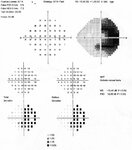 |
Chiasmal Compression - Vascular - OCSOD1 | 2024-07 | Image | ehsl_novel_nrm |
| 105 |
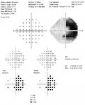 |
Chiasmal Compression - Vascular - OCSOD2 | 2024-07 | Image | ehsl_novel_nrm |
| 106 |
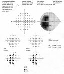 |
Chiasmal Compression - Vascular - OCSOS1 | 2024-07 | Image | ehsl_novel_nrm |
| 107 |
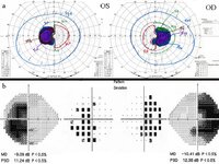 |
Chiasmal Compression - Vascular - VF | 2024-07 | Image | ehsl_novel_nrm |
| 108 |
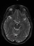 |
Chiasmal Compression by a Vascular Loop | 2024-07 | Image | ehsl_novel_nrm |
| 109 |
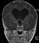 |
Chiasmal Compression from a Dilated Third Ventricle in a Patient with Severe Hydrocephalus (MRI) | 2024-07 | Image | ehsl_novel_nrm |
| 110 |
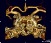 |
Chiasmal Compression from a Vascular Loop | 2024-07 | Image | ehsl_novel_nrm |
| 111 |
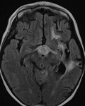 |
Chiasmal PNET - 3 | 2024-07 | Image | ehsl_novel_nrm |
| 112 |
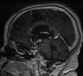 |
Chiasmal PNET - 8 | 2024-07 | Image | ehsl_novel_nrm |
| 113 |
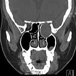 |
Cholesterol Granuloma - 08 | 2024-07 | Image | ehsl_novel_nrm |
| 114 |
 |
Choriocarcinoma Primary - 1a | 2024-07 | Image | ehsl_novel_nrm |
| 115 |
 |
Choriocarcinoma Primary - 1b | 2024-07 | Image | ehsl_novel_nrm |
| 116 |
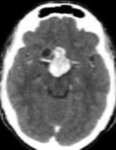 |
Choriocarcinoma Primary - 1c | 2024-07 | Image | ehsl_novel_nrm |
| 117 |
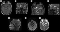 |
Choriocarcinoma Primary - 2 | 2024-07 | Image | ehsl_novel_nrm |
| 118 |
 |
Choriocarcinoma Primary - 2a | 2024-07 | Image | ehsl_novel_nrm |
| 119 |
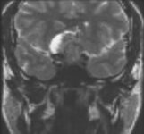 |
Choriocarcinoma Primary - 2b | 2024-07 | Image | ehsl_novel_nrm |
| 120 |
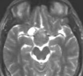 |
Choriocarcinoma Primary - 2c | 2024-07 | Image | ehsl_novel_nrm |
| 121 |
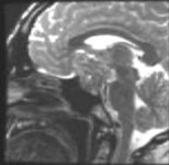 |
Choriocarcinoma Primary - 2d | 2024-07 | Image | ehsl_novel_nrm |
| 122 |
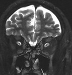 |
Choroidal Folds - 3 | 2024-07 | Image | ehsl_novel_nrm |
| 123 |
 |
Choroidal Hypoperfusion in a Patient with CADASIL | 2024-07-10 | Image | ehsl_novel_nrm |
| 124 |
 |
Clinical Features of Periopitic Neuritis | 1999-03-15 | Text | ehsl_novel_nam |
| 125 |
 |
Clinical Use of the Multifocal Electroretinogram (MERG) in Neuro-Ophthalmology: Distinguishing Optic Nerve Disease from Retinal Disfunction | 1999-03-14 | Text | ehsl_novel_nam |
| 126 |
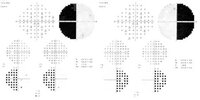 |
Complete Bitemporal Hemianopia from Chiasmal Damage | 2024-07 | Image | ehsl_novel_nrm |
| 127 |
 |
Conopeptide discovery through calcium imaging | 2015 | Text | ir_htca |
| 128 |
 |
CRANIO - Not23 - Lowgrade_Glioma - 3rd Ventricle - 6-12-23 | 2023-06-12 | Image | ehsl_novel_nrm |
| 129 |
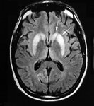 |
Creuzfeldt-Jakob Disease - FLAIR sCJD - Arrows | 2024-07 | Image | ehsl_novel_nrm |
| 130 |
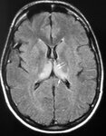 |
Creuzfeldt-Jakob Disease - FLAIR vCJD - Arrows | 2024-07 | Image | ehsl_novel_nrm |
| 131 |
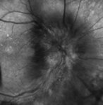 |
Cytomegalovirus - Optic Neuritis - 1bw | 2024-07 | Image | ehsl_novel_nrm |
| 132 |
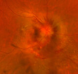 |
Cytomegalovirus - Optic Neuritis - 1c | 2024-07 | Image | ehsl_novel_nrm |
| 133 |
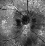 |
Cytomegalovirus - Optic Neuritis - 2bw | 2024-07 | Image | ehsl_novel_nrm |
| 134 |
 |
Cytomegalovirus - Optic Neuritis - 2c | 2024-07 | Image | ehsl_novel_nrm |
| 135 |
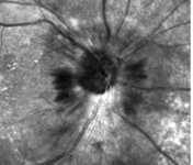 |
Cytomegalovirus - Optic Neuritis - 3bw | 2024-07 | Image | ehsl_novel_nrm |
| 136 |
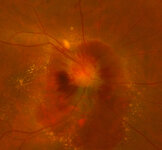 |
Cytomegalovirus - Optic Neuritis - 3c | 2024-07 | Image | ehsl_novel_nrm |
| 137 |
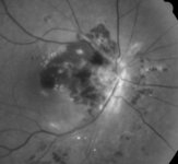 |
Cytomegalovirus - Optic Neuritis - 4bw | 2024-07 | Image | ehsl_novel_nrm |
| 138 |
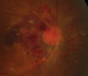 |
Cytomegalovirus - Optic Neuritis - 4c | 2024-07 | Image | ehsl_novel_nrm |
| 139 |
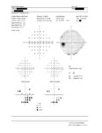 |
Cytomegalovirus - Optic Neuritis - in Left Eye | 2024-07 | Image | ehsl_novel_nrm |
| 140 |
 |
Development of contrast enhancement after long-term observation of a dysembryoplastic neuroepithelial tumor | 2006 | Text | ir_uspace |
| 141 |
 |
DWI - Diffusion Weighted Imaging | 2019-02 | Image/MovingImage | ehsl_novel_lee |
| 142 |
 |
Family Album Tomography | 2021-06 | Image/MovingImage | ehsl_novel_lee |
| 143 |
 |
Fluorescein Angiography (F A) Abnormalities in Vasculitis Related, Unilateral Ischemic Optic Neuropathy (AION) | 1999-03-15 | Text | ehsl_novel_nam |
| 144 |
 |
The Future of Neuroimaging | 2008-03-13 | Text | ehsl_novel_nam |
| 145 |
 |
Generalized diffraction-stack migration | 2012-12 | Text | ir_etd |
| 146 |
 |
Hands On, Hands Off OCT (video) | 2016-02-29 | Image/MovingImage | ehsl_novel_nam |
| 147 |
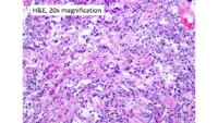 |
Histopathology and Immunohistochemistry of a Low-grade Glioma Thought on Imaging to Be a Craniopharyngioma | | Image | ehsl_novel_nrm |
| 148 |
 |
Homonymous Visual Field Defects in Patients Without Structural Lesions on Neuroimaging | 1999-03-15 | Text | ehsl_novel_nam |
| 149 |
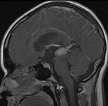 |
Hydrocephalus Caused by a Large Pineal Region Mass | 2024-07 | Image | ehsl_novel_nrm |
| 150 |
 |
Hydrocephalus Causing Downward Compression and Displacement of the Optic Chiasm | 2024-07 | Image | ehsl_novel_nrm |
| 151 |
 |
Hydrocephalus Causing Downward Compression of the Optic Chiasm by an Enlarged Third Ventricle | 2024-07 | Image | ehsl_novel_nrm |
| 152 |
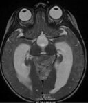 |
Hydrocephalus from a Large Posterior Fossa Mass | 2024-07 | Image | ehsl_novel_nrm |
| 153 |
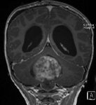 |
Hydrocephalus from Large Posterior Fossa Mass | 2024-07 | Image | ehsl_novel_nrm |
| 154 |
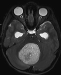 |
Hydrocephalus from Large Posterior Fossa Mass | 2024-07 | Image | ehsl_novel_nrm |
| 155 |
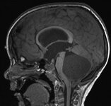 |
Hydrocephalus from Large Posterior Fossa Mass | 2024-07 | Image | ehsl_novel_nrm |
| 156 |
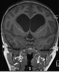 |
Hyrocephalus with Downward Compression of the Optic Chiasm by an Enlarged Third Ventricle | 2024-07 | Image | ehsl_novel_nrm |
| 157 |
 |
Imaging Disease of the Chiasm | 2023-03 | Image/MovingImage | ehsl_novel_nam |
| 158 |
 |
Imaging of a Suprasellar Lesion Thought to Be a Craniopharyngioma But on Biopsy was a Low-grade Glioma | | Image | ehsl_novel_nrm |
| 159 |
 |
Imaging of the Cavernous Sinus and Skull Base | 2008-03-13 | Text | ehsl_novel_nam |
| 160 |
 |
Imaging the Nerve Fiber Layer and Optic Disc | 2001-02-21 | Text | ehsl_novel_nam |
| 161 |
 |
Indocyanine Green Angiography Findings in Acute Idiopathic Blind Spot Enlargement Syndrome | 1999-03-15 | Text | ehsl_novel_nam |
| 162 |
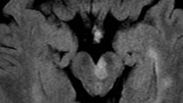 |
Infarction Shown on Diffusion Weighted MRI | 2021-09 | Image/MovingImage | ehsl_novel_jdt |
| 163 |
 |
Inter-Rater Reliability in the New Low-Contrast Sloan Letter Charts in Patients with Multiple Sclerosis | 1999-03-14 | Text | ehsl_novel_nam |
| 164 |
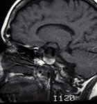 |
Intracavernous Aneurysm (MRI) | 2024-07-10 | Image | ehsl_novel_nrm |
| 165 |
 |
Intracranial Cavernoma | 2024-07-10 | Image | ehsl_novel_nrm |
| 166 |
 |
Intracranial Cavernoma | 2024-07-10 | Image | ehsl_novel_nrm |
| 167 |
 |
Intracranial Cavernoma | 2024-07-10 | Image | ehsl_novel_nrm |
| 168 |
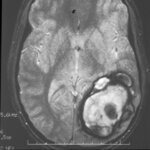 |
Intracranial Cavernoma | 2024-07-10 | Image | ehsl_novel_nrm |
| 169 |
 |
Intracranial Cavernoma | 2024-07-10 | Image | ehsl_novel_nrm |
| 170 |
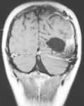 |
Intracranial Cavernoma | 2024-07-10 | Image | ehsl_novel_nrm |
| 171 |
 |
Intracranial Cavernoma | 2024-07-10 | Image | ehsl_novel_nrm |
| 172 |
 |
Intracranial Cavernoma | 2024-07-10 | Image | ehsl_novel_nrm |
| 173 |
 |
Introduction to Neuroimaging in Neuro-Ophthalmology | 2018-08 | Text | ehsl_novel_novel |
| 174 |
 |
Invited Commentary: Evaluation of Horner Syndrome in the MRI Era | 2018-03 | Text | ehsl_novel_jno |
| 175 |
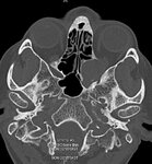 |
Large Left Orbital and Retro-orbital Cholesterol Granuloma | 2024-07 | Image | ehsl_novel_nrm |
| 176 |
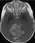 |
Large Posterior Fossa Mass with Brainstem Compression | 2024-07 | Image | ehsl_novel_nrm |
| 177 |
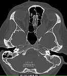 |
Left Orbital and Retro-orbital Cholesterol Granuloma | 2024-07 | Image | ehsl_novel_nrm |
| 178 |
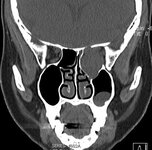 |
Left Orbital Invasion by a Large Cholesterol Granuloma | 2024-07 | Image | ehsl_novel_nrm |
| 179 |
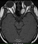 |
Left Orbital Invasion by Cholesterol Granuloma | 2024-07 | Image | ehsl_novel_nrm |
| 180 |
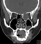 |
Left Orbital Invasion by Large Cholesterol Granuloma | 2024-07 | Image | ehsl_novel_nrm |
| 181 |
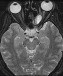 |
Left Orbital Invasion by Large Cholesterol Granuloma | 2024-07 | Image | ehsl_novel_nrm |
| 182 |
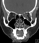 |
Left orbital invasion by large cholesterol granuloma | 2024-07 | Image | ehsl_novel_nrm |
| 183 |
 |
Left-sided Intracavernous Aneurysm | 2024-07-10 | Image | ehsl_novel_nrm |
| 184 |
 |
Limitations of Neuroimaging | 2008-03-13 | Text | ehsl_novel_nam |
| 185 |
 |
Magnetic Resonance Imaging of Superior Oblique Muscle Atrophy in Trochlear Nerve Schwannoma | 2013-02-12 | Text | ehsl_novel_nam |
| 186 |
 |
Magnetic Resonance Imaging of the Human Lateral Geniculate Body | 1990-02-04 | Text | ehsl_novel_nam |
| 187 |
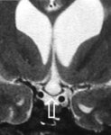 |
Marked Compression and Downward Displacement of the Chiasm by an Enlarged Third Ventricle in a Patient with Hydrocephalus | 2024-07 | Image | ehsl_novel_nrm |
| 188 |
 |
Metamaterials and their applications in imaging | 2017 | Text | ir_etd |
| 189 |
 |
Modern Imaging of Optic Disc Drusen | 2021 | Text | ehsl_novel_novel |
| 190 |
 |
MR Imaging of the Cavernous Sinus | 1990-02-07 | Text | ehsl_novel_nam |
| 191 |
 |
MRI Diagnosis of Clival Cancer and Sixth Nerve Palsy | 2023-03 | Text | ehsl_novel_jno |
| 192 |
 |
MRI Findings in Giant Cell Arteritis | 2022 | Text | ehsl_novel_eec |
| 193 |
 |
MRI in Neuro-ophthalmology | 2019-03 | Image/MovingImage | ehsl_novel_lee |
| 194 |
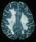 |
MRI Showing Extensive White Matter Disease in a Patient with CADASIL | 2024-07-10 | Image | ehsl_novel_nrm |
| 195 |
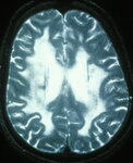 |
MRI Showing Extensive White Matter Disease in a Patient with CADASIL | 2024-07-10 | Image | ehsl_novel_nrm |
| 196 |
 |
MRI T1, Non-contrast Sagittal Image of Large Parietal-occipital Cavernoma | 2024-07-10 | Image | ehsl_novel_nrm |
| 197 |
 |
MRI, Axial T2 Image, Showing Large Left Parietal-occipital Cavernoma | 2024-07-10 | Image | ehsl_novel_nrm |
| 198 |
 |
MRI, T1 Axial, Following Gadolinium Administration, Showing Large, Left Parietal-occipital Cavernoma | 2024-07-10 | Image | ehsl_novel_nrm |
| 199 |
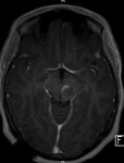 |
MRI, T1 Image After Contrast Injection, Shows a Lesion in the Midbrain that Caused a Benedikt Syndrome | 2024-07-10 | Image | ehsl_novel_nrm |
| 200 |
 |
MRI, T2 Axial Image, Showing Midbrain Lesion Causing a Benedikt Syndrome | 2024-07-10 | Image | ehsl_novel_nrm |