|
|
Title | Date | Type | Setname |
| 1 |
 |
Ambati, Balamurali K., M.D., Ph.D., M.B.A. | | Image | ehsl_hhs |
| 2 |
 |
Anatomical Crucifixion | | Image | uu_aah_art |
| 3 |
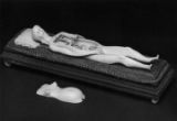 |
Anatomical Figure of a Pregnant Woman with removable parts (view 1) | | Image | uu_aah_art |
| 4 |
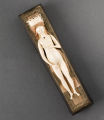 |
Anatomical Figure of a Pregnant Woman with removable parts (view 2) | | Image | uu_aah_art |
| 5 |
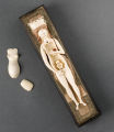 |
Anatomical Figure of a Pregnant Woman with removable parts (view 3) | | Image | uu_aah_art |
| 6 |
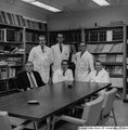 |
Anatomy Department (1970) | | Image | ehsl_hhs |
| 7 |
 |
Anatomy of a Seated Woman | | Image | uu_aah_art |
| 8 |
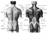 |
Back View of Musculature and Skeleton | | Image | uu_aah_art |
| 9 |
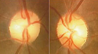 |
Bilateral Band Atrophy of Optic Nerves from Optic Chiasmal Compression | 2024-07 | Image | ehsl_novel_nrm |
| 10 |
 |
Bones of the Hand | | Image | uu_aah_art |
| 11 |
 |
The Bones of the Human Body | | Image | uu_aah_art |
| 12 |
 |
Callister, A. Cyril, M.D. | 1970 | Image | ehsl_hhs |
| 13 |
 |
Callister, A. Cyril, M.D. | | Image | ehsl_hhs |
| 14 |
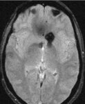 |
Cavernoma in Region of Chiasm (SWI MRI) | 2024-07 | Image | ehsl_novel_nrm |
| 15 |
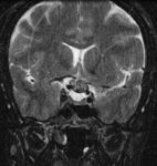 |
Cavernoma of the Optic Chiasm Causing Chiasmal Apoplexy | 2024-07 | Image | ehsl_novel_nrm |
| 16 |
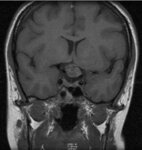 |
Cavernoma of the Optic Chiasm Causing Chiasmal Apoplexy | 2024-07 | Image | ehsl_novel_nrm |
| 17 |
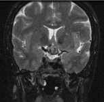 |
Cavernoma of the Optic Chiasm Causing Chiasmal Apoplexy | 2024-07 | Image | ehsl_novel_nrm |
| 18 |
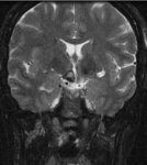 |
Cavernoma of the Optic Chiasm Causing Chiasmal Apoplexy | 2024-07 | Image | ehsl_novel_nrm |
| 19 |
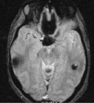 |
Cavernoma of the Optic Chiasm Causing Chiasmal Apoplexy | 2024-07 | Image | ehsl_novel_nrm |
| 20 |
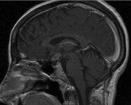 |
Cavernoma of the Optic Chiasm Causing Chiasmal Apoplexy | 2024-07 | Image | ehsl_novel_nrm |
| 21 |
 |
Chess Player, back view | | Image | uu_aah_art |
| 22 |
 |
Chess Player, front view | | Image | uu_aah_art |
| 23 |
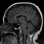 |
Chiari Malformation - 01 | 2024-07 | Image | ehsl_novel_nrm |
| 24 |
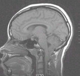 |
Chiari Malformation - 02 | 2024-07 | Image | ehsl_novel_nrm |
| 25 |
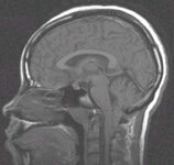 |
Chiari Malformation - 03 | 2024-07 | Image | ehsl_novel_nrm |
| 26 |
 |
Chiari Malformation - 04 | 2024-07 | Image | ehsl_novel_nrm |
| 27 |
 |
Chiari Malformation - 05 | 2024-07 | Image | ehsl_novel_nrm |
| 28 |
 |
Chiari Malformation - 06 | 2024-07 | Image | ehsl_novel_nrm |
| 29 |
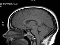 |
Chiari Malformation - a | 2024-07 | Image | ehsl_novel_nrm |
| 30 |
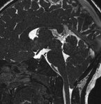 |
Chiari Malformation in a Patient who Presented with Papilledema (MRI) | 2024-07 | Image | ehsl_novel_nrm |
| 31 |
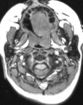 |
Chiari Malformation in a Patient who Presented with Papilledema (MRI) | 2024-07 | Image | ehsl_novel_nrm |
| 32 |
 |
Chiari Malformation in a Patient who Presented with Papilledema (MRI) | 2024-07 | Image | ehsl_novel_nrm |
| 33 |
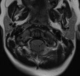 |
Chiari Malformation with Papilledema - 02 | 2024-07 | Image | ehsl_novel_nrm |
| 34 |
 |
Chiari Malformation with Papilledema - OD | 2024-07 | Image | ehsl_novel_nrm |
| 35 |
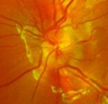 |
Chiari Malformation with Papilledema - OS | 2024-07 | Image | ehsl_novel_nrm |
| 36 |
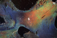 |
Chiasm Anatomy - 001 | 2024-07 | Image | ehsl_novel_nrm |
| 37 |
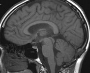 |
Chiasm Anatomy - Birdbeak | 2024-07 | Image | ehsl_novel_nrm |
| 38 |
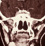 |
Chiasm Anatomy - Chiasmal Adenohypophysis - 1 | 2024-07 | Image | ehsl_novel_nrm |
| 39 |
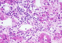 |
Chiasm Anatomy - Chiasmal Adenohypophysis - 2 | 2024-07 | Image | ehsl_novel_nrm |
| 40 |
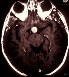 |
Chiasm Anatomy - Chiasmal Germinoma - 1 | 2024-07 | Image | ehsl_novel_nrm |
| 41 |
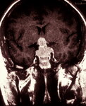 |
Chiasm Anatomy - Chiasmal Germinoma - 2 | 2024-07 | Image | ehsl_novel_nrm |
| 42 |
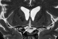 |
Chiasm Anatomy - Chiasmal Infarction | 2024-07 | Image | ehsl_novel_nrm |
| 43 |
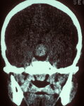 |
Chiasm Anatomy - Germinoma - 01 | 2024-07 | Image | ehsl_novel_nrm |
| 44 |
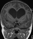 |
Chiasm Anatomy - Hydrocephalus - 002 | 2024-07 | Image | ehsl_novel_nrm |
| 45 |
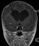 |
Chiasm Anatomy - Hydrocephalus - 006 | 2024-07 | Image | ehsl_novel_nrm |
| 46 |
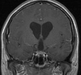 |
Chiasm Anatomy - Hydrocephalus - 02 | 2024-07 | Image | ehsl_novel_nrm |
| 47 |
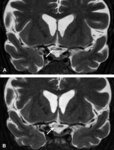 |
Chiasm Anatomy - Infarction - 001 | 2024-07 | Image | ehsl_novel_nrm |
| 48 |
 |
Chiasm Anatomy - Infarction - 002 | 2024-07 | Image | ehsl_novel_nrm |
| 49 |
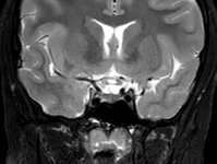 |
Chiasm Anatomy - Infarction - 01 | 2024-07 | Image | ehsl_novel_nrm |
| 50 |
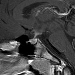 |
Chiasm Anatomy - Infarction - 03 | 2024-07 | Image | ehsl_novel_nrm |
| 51 |
 |
Chiasm Anatomy - Infarction - 04 | 2024-07 | Image | ehsl_novel_nrm |
| 52 |
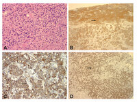 |
Chiasm Anatomy - Pit Carcinoma 2 | 2024-07 | Image | ehsl_novel_nrm |
| 53 |
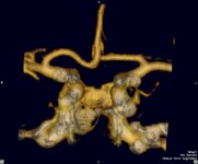 |
Chiasmal Compression - Vascular - 010 | 2024-07 | Image | ehsl_novel_nrm |
| 54 |
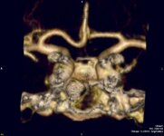 |
Chiasmal Compression - Vascular - 08 | 2024-07 | Image | ehsl_novel_nrm |
| 55 |
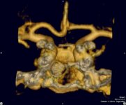 |
Chiasmal Compression - Vascular - 09 | 2024-07 | Image | ehsl_novel_nrm |
| 56 |
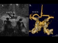 |
Chiasmal Compression - Vascular - CTA | 2024-07 | Image | ehsl_novel_nrm |
| 57 |
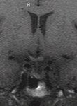 |
Chiasmal Compression - Vascular - g | 2024-07 | Image | ehsl_novel_nrm |
| 58 |
 |
Chiasmal Compression - Vascular - h | 2024-07 | Image | ehsl_novel_nrm |
| 59 |
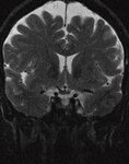 |
Chiasmal Compression - Vascular - i | 2024-07 | Image | ehsl_novel_nrm |
| 60 |
 |
Chiasmal Compression - Vascular - j | 2024-07 | Image | ehsl_novel_nrm |
| 61 |
 |
Chiasmal Compression - Vascular - k | 2024-07 | Image | ehsl_novel_nrm |
| 62 |
 |
Chiasmal Compression - Vascular - KineticOD | 2024-07 | Image | ehsl_novel_nrm |
| 63 |
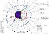 |
Chiasmal Compression - Vascular - KineticOS | 2024-07 | Image | ehsl_novel_nrm |
| 64 |
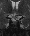 |
Chiasmal Compression - Vascular - l | 2024-07 | Image | ehsl_novel_nrm |
| 65 |
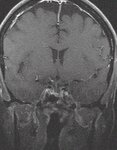 |
Chiasmal Compression - Vascular - m | 2024-07 | Image | ehsl_novel_nrm |
| 66 |
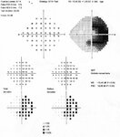 |
Chiasmal Compression - Vascular - OCSOD1 | 2024-07 | Image | ehsl_novel_nrm |
| 67 |
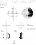 |
Chiasmal Compression - Vascular - OCSOD2 | 2024-07 | Image | ehsl_novel_nrm |
| 68 |
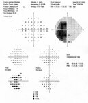 |
Chiasmal Compression - Vascular - OCSOS1 | 2024-07 | Image | ehsl_novel_nrm |
| 69 |
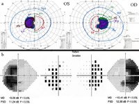 |
Chiasmal Compression - Vascular - VF | 2024-07 | Image | ehsl_novel_nrm |
| 70 |
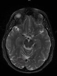 |
Chiasmal Compression by a Vascular Loop | 2024-07 | Image | ehsl_novel_nrm |
| 71 |
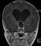 |
Chiasmal Compression from a Dilated Third Ventricle in a Patient with Severe Hydrocephalus (MRI) | 2024-07 | Image | ehsl_novel_nrm |
| 72 |
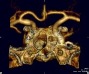 |
Chiasmal Compression from a Vascular Loop | 2024-07 | Image | ehsl_novel_nrm |
| 73 |
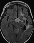 |
Chiasmal PNET - 3 | 2024-07 | Image | ehsl_novel_nrm |
| 74 |
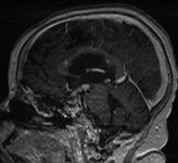 |
Chiasmal PNET - 8 | 2024-07 | Image | ehsl_novel_nrm |
| 75 |
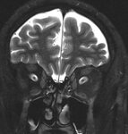 |
Choroidal Folds - 3 | 2024-07 | Image | ehsl_novel_nrm |
| 76 |
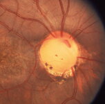 |
Coloboma - 001 | 2024-07 | Image | ehsl_novel_nrm |
| 77 |
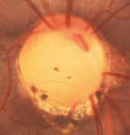 |
Coloboma - 002 | 2024-07 | Image | ehsl_novel_nrm |
| 78 |
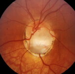 |
Coloboma - 003 | 2024-07 | Image | ehsl_novel_nrm |
| 79 |
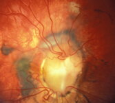 |
Coloboma - 004 | 2024-07 | Image | ehsl_novel_nrm |
| 80 |
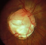 |
Coloboma - 005 | 2024-07 | Image | ehsl_novel_nrm |
| 81 |
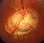 |
Coloboma - 007 | 2024-07 | Image | ehsl_novel_nrm |
| 82 |
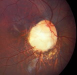 |
Coloboma - 009 | 2024-07 | Image | ehsl_novel_nrm |
| 83 |
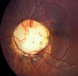 |
Coloboma - 010 | 2024-07 | Image | ehsl_novel_nrm |
| 84 |
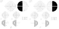 |
Complete Bitemporal Hemianopia from Chiasmal Damage | 2024-07 | Image | ehsl_novel_nrm |
| 85 |
 |
Diagram of Bent Leg, Outside View | | Image | uu_aah_art |
| 86 |
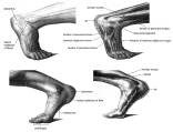 |
Diagram of Flexed Foot, outside and inside views | | Image | uu_aah_art |
| 87 |
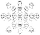 |
Diagram of Heads in Perspective | | Image | uu_aah_art |
| 88 |
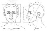 |
Diagram of Proportions of the Head, front and profile views | | Image | uu_aah_art |
| 89 |
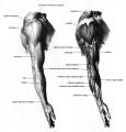 |
Diagram of Skeletal and Muscular Structure of Outer Arm | | Image | uu_aah_art |
| 90 |
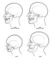 |
Diagram of Skulls at Various Stages of Aging | | Image | uu_aah_art |
| 91 |
 |
Diagrams of the Musculature and Skeletal Structure of the Head and Neck, front and side views | | Image | uu_aah_art |
| 92 |
 |
Dougherty, Thomas F., Ph.D. | 1964 | Image | ehsl_hhs |
| 93 |
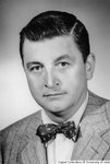 |
Dougherty, Thomas F., Ph.D. (1947) | | Image | ehsl_hhs |
| 94 |
 |
Dougherty, Thomas F., Ph.D. (1965) | | Image | ehsl_hhs |
| 95 |
 |
Drs. Samuels, Nelson & Dougherty | | Image | ehsl_hhs |
| 96 |
 |
Ephraim G. Gowans | | Image | dha_cp |
| 97 |
 |
Fragmented Plastinate | | Image | uu_aah_art |
| 98 |
 |
Freudenberger, Clay B., M.D. Ph.D. | | Image | ehsl_hhs |
| 99 |
 |
Freudenberger, Clay B., M.D. Ph.D. | | Image | ehsl_hhs |
| 100 |
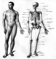 |
Front View of Male Figure and Skeleton | | Image | uu_aah_art |