TO
Filters: Collection: ehsl_novel_* Type: "Text"
| Title | Curriculum | Description | Subject | Collection | ||
|---|---|---|---|---|---|---|
| 1 |
 |
Progression Over 5 Years of Prelaminar Hyperreflective Lines to Optic Disc Drusen in the Copenhagen Child Cohort 2000 Eye Study | IC-D1hvi-optic-nerve-drusen | Child; Denmark / epidemiology; Female; Humans; Incidence; Male; Optic Disk / diagnostic imaging; Optic Disk Drusen / diagnosis; Optic Disk Drusen / epidemiology; Prospective Studies; Retinal Ganglion Cells / pathology; Tomography, Optical Coherence / methods; Visual Acuity; Visual Fields / physiolog... | Neuro-Ophthalmology Virtual Education Library - Journal of Neuro-Ophthalmology Archives: https://novel.utah.edu/jno/ | |
| 2 |
 |
Optic Disc Drusen | Optic disc drusen are abnormal deposits of protein-like material in the optic disc - the front part of the optic nerve. Updated April 2020. | Optic Disc Drusen; Patient Brochure | Neuro-ophthalmology Virtual Education Library: NOVEL http://NOVEL.utah.edu | |
| 3 |
 |
Focal Capillary Dropout Associated With Optic Disc Drusen Using Optical Coherence Tomographic Angiography | Capillaries; Female; Fluorescein Angiography; Fundus Oculi; Humans; Middle Older people; Nerve Fibers; Optic Disk; Optic Disk Drusen; Retinal Ganglion Cells; Retinal Vessels; Scotoma; Tomography, Optical Coherence; Visual Acuity; Visual Fields | Neuro-Ophthalmology Virtual Education Library - Journal of Neuro-Ophthalmology Archives: https://novel.utah.edu/jno/ | ||
| 4 |
 |
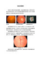
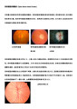
Visible Optic Disc Drusen in a Children | Optic disc drusen are acellular deposits located in the head of the optic nerve that increase in size and become calcified over time. Drusen tend to buried in early life and become superficial and visible later in childhood, at an average age of 10-12 years. We had the opportunity to evaluate a 5-ye... | Pediatric Neuro-ophthalmology; Diagnostic Tests (ERG, VER, OCT, HRT, mfERG, etc); Miscellaneous | Neuro-ophthalmology Virtual Education Library: NOVEL http://NOVEL.utah.edu | |
| 5 |
 |
Modern Imaging of Optic Disc Drusen | IC-C6bvi-optic-disc-drusen; IC-D1hvi-optic-nerve-drusen | This is a short powerpoint describing imaging techniques (specifically OCT-EDI, fundus autofluorescence, and B-scan ultrasonography) for optic disc drusen. Examples of these techniques are included. | Optic Disc Drusen; Imaging; OCT-EDI; Fundus Autofluorescence; B-scan Ultrasonography | Neuro-ophthalmology Virtual Education Library: NOVEL http://NOVEL.utah.edu |
| 6 |
 |
Optic Disc Drusen: Insights on Diagnosis | Guidelines for enhanced-depth imaging have improved the diagnosis of optic disc drusen and attempt to address confusion within the literature. In particular the presence of peripapillary mass-like structures have caused this confusion and potentially lead to misclassification of some disc oedema. Th... | Optic Disc Drusen; Peripapillary Hyperreflective Ovoid Mass-like Structures; Optical Coherence Tomography; Ocular Ultrasound; Visual Field | Neuro-ophthalmology Virtual Education Library: NOVEL http://NOVEL.utah.edu | |
| 7 |
 |
The Influence of Volume and Anatomic Location of Optic Disc Drusen on Sensitivity of Autofluorescence | Optic disc drusen are acellular deposits in the optic nerve head. Optic disc drusen can be diagnosed using different imaging modalities such as enhanced depth imaging optical coherence tomography and autofluorescence. It is unknown which factors determine the sensitivity of autofluorescence. The aim... | Diagnostic Tests (ERG, VER, OCT, HRT, mfERG, etc); Eyelid & Adnexal Disease | Neuro-ophthalmology Virtual Education Library: NOVEL http://NOVEL.utah.edu | |
| 8 |
 |
Walsh & Hoyt: Pseudopapilledema Associated with Optic Disc Drusen | IC-C6bvi-optic-disc-drusen | The word drusen, of Germanic origin, originally meant tumor, swelling, or tumescence. According to Lorentzen, the word was used in the mining industry approximately 500 years ago to indicate a crystal-filled space in a rock. Drusen of the optic disc were first described clinically by Liebreich in 18... | Eye Abnormalities; Pseudopapilledema; Optic Disc Drusen; Buried Drusen; Optic Disc Anomalies; Congenital Blurred Disc; Congenital Optic Nerve Anomalies | Neuro-ophthalmology Virtual Education Library: NOVEL http://NOVEL.utah.edu |
| 9 |
 |
OCT and Optic Nerve Head Drusen (abstract) | Patient Care; Medical Knowledge; PBLI; VBnflaopticalcoherencetomography; KBDexposeddrusen; KBDburieddrusen; KBDlittlereddiscs; KBDcongenitalblurreddisc; IC-C6bvi-optic-disc-drusen | Optic nerve head drusen (ONHD) are deposits of calcium, amino and nucleic acids, and mucopolysaccharides, which may be buried within the optic nerve or lie at the surface of the optic disc.1 These deposits often become calcified over time. Clinically apparent ONHD are estimated to occur in 0.3% of t... | Spectral-Domain OCT; Superficial ONHD; Buried ONHD; Enhanced Depth Imaging OCT; Swept Source OCT; Papilledema; Pseudopapilledema | Neuro-ophthalmology Virtual Education Library: NOVEL http://NOVEL.utah.edu |
| 10 |
 |
In-Clinic Detection of Optic Disc Drusen Using Hand-Held Ultrasound Technology | Differentiation of pseudopapilledema from true papilledema is a common reason for referral to Neuro-ophthalmology. Optic disc drusen is a frequent cause of pseudopapilledema, though buried drusen may not be visible on fundus examination. Ancillary testing including ultrasonography can rapidly identi... | Diagnostic Tests (ERG, VER, OCT, HRT, mfERG, etc); Orbit/Ocular Pathology | Neuro-ophthalmology Virtual Education Library: NOVEL http://NOVEL.utah.edu | |
| 11 |
 |
Optic Disc Drusen Visualization with Auto-Fluorescent Imaging | Minimally buried optic disc drusen may present as swelling of the optic disc. Drusen auto-fluoresce when excited by short wavelength light. To distinguish disc edema from buried disc drusen, auto-fluorescent imaging may be useful. | Optic Disc Drusen; Auto-Fluorescence; Scanning Laser Ophthalmoscope | Neuro-ophthalmology Virtual Education Library: NOVEL http://NOVEL.utah.edu | |
| 12 |
 |
Optic Disc Drusen (Simplified Chinese) | Optic disc drusen are abnormal deposits of protein-like material in the optic disc - the front part of the optic nerve. | Optic Disc Drusen; Patient Brochure | Neuro-ophthalmology Virtual Education Library: NOVEL http://NOVEL.utah.edu | |
| 13 |
 |
Optic Disc Drusen (Traditional Chinese) | Optic disc drusen are abnormal deposits of protein-like material in the optic disc - the front part of the optic nerve. | Optic Disc Drusen; Patient Brochure | Neuro-ophthalmology Virtual Education Library: NOVEL http://NOVEL.utah.edu | |
| 14 |
 |
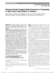
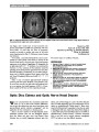
Enhanced Depth Imaging Optical Coherence Tomography Technology Reveals a Significant Association Between Optic Nerve Drusen Anterior Displacement and Retinal Nerve Fiber Layer Thinning Over Time | Optic Disc Drusen; OCT; EDI | Neuro-Ophthalmology Virtual Education Library - Journal of Neuro-Ophthalmology Archives: https://novel.utah.edu/jno/ | ||
| 15 |
 |
Enhanced Depth Imaging Optical Coherence Tomography of Optic Nerve Head Drusen in Children | Adolescent; Child; Cross-Sectional Studies; Female; Humans; Male; Nerve Fibers / pathology; Optic Disk / diagnostic imaging; Optic Disk Drusen / diagnosis; Optic Disk Drusen / physiopathology; Retinal Ganglion Cells / pathology; Retrospective Studies; Tomography, Optical Coherence / methods; Visual ... | Neuro-Ophthalmology Virtual Education Library - Journal of Neuro-Ophthalmology Archives: https://novel.utah.edu/jno/ | ||
| 16 |
 |
Exposed Drusen (PowerPoint) | curriculum_fellow; KBDexposeddrusen; KBDdcaexposeddrusen; KBDdcapseudoexposeddrusen | PP25a: Left eye: Severe visual field defect. PP25b: right eye with exposed drusen and field loss: visual field defects; PP25c: right eye visual field PP25d: left eye visual field. | Pseudopapilledema; Exposed Drusen | Neuro-ophthalmology Virtual Education Library: NOVEL http://NOVEL.utah.edu |
| 17 |
 |
Optic Disc Edema and Optic Nerve Head Drusen | Female; Humans; Male; Nerve Fibers / diagnostic imaging; Optic Disk / diagnostic imaging; Optic Disk Drusen / complications; Papilledema / etiology; Radiography; Tomography, Optical Coherence | Neuro-Ophthalmology Virtual Education Library - Journal of Neuro-Ophthalmology Archives: https://novel.utah.edu/jno/ | ||
| 18 |
 |
Concomitant Optic Disc Drusen and Papilledema Due to Idiopathic Intracranial Hypertension in a Pediatric Cohort | Recent studies have shown higher rates of optic disc drusen (ODD) among pediatric patients with idiopathic intracranial hypertension (IIH). In this study, we retrospectively reviewed the patients with confirmed ODD to evaluate the rate and characteristics of concomitant IIH. | Pediatric Neuro-ophthalmology; Pseudotumor Cerebri | Neuro-ophthalmology Virtual Education Library: NOVEL http://NOVEL.utah.edu | |
| 19 |
 |
Measurement of Scleral Canal Area Using Optical Coherence Tomography in Patients with Optic Nerve Drusen | The pathophysiology of optic nerve drusen (OND) is speculative. It has been proposed that optic nerve drusen are formed when retinal ganglion cell axons die and extrude their mitochondria (Tso, 1981). These extruded mitochondria serve as nidifor calcification. However, it is not clear why these gang... | Optic Nerve Drusen; Optical Coherence Tomography; Scleral Canal | Neuro-ophthalmology Virtual Education Library: NOVEL http://NOVEL.utah.edu | |
| 20 |
 |
Peripapillary Hyperreflective Ovoid Mass-Like Structures: Is It Optic Disc Drusen or Not?: Response | Humans, Optic Disk, Optic Disk Drusen, Papilledema, Tomography, Optical Coherence | Neuro-Ophthalmology Virtual Education Library - Journal of Neuro-Ophthalmology Archives: https://novel.utah.edu/jno/ | ||
| 21 |
 |
Scanning Laser Polarimetry for the Detection of Nerve Fiber Layer Defects in Patients with Optic Nerve Drusen | Patients with optic nerve drusen may have other diseases of the optic nerve, including glaucoma, complicating the evaluation of visual field defects. In diseases of the optic nerve, visual field defects result from loss of ganglion cells and thinning of the retinal nerve fiber layer. We performed sc... | Optic Nerve Drusen; Visual Field Defects; Nerve Fiber Layer Defects; Laser Polarimetry | Neuro-ophthalmology Virtual Education Library: NOVEL http://NOVEL.utah.edu | |
| 22 |
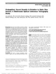 |
Peripapillary Vessel Density in Relation to Optic Disc Drusen: A Multimodal Optical Coherence Tomography Study | ODD: Visual Field Defects; Optical Coherence Tomography | Neuro-Ophthalmology Virtual Education Library - Journal of Neuro-Ophthalmology Archives: https://novel.utah.edu/jno/ | ||
| 23 |
 |
Peripapillary Hyperreflective Ovoid Mass-Like Structures: Is It Optic Disc Drusen or Not? | Humans, Optic Disk, Optic Disk Drusen, Papilledema, Tomography, Optical Coherence | Neuro-Ophthalmology Virtual Education Library - Journal of Neuro-Ophthalmology Archives: https://novel.utah.edu/jno/ | ||
| 24 |
 |
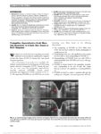
Novel Cellular and Animal Models of Optic Disc Drusen | Optic disc drusen (ODD) are calcified deposits of the anterior optic nerve and result in visual field loss in majority of patients.1 Most common cause of vision loss is anterior ischemic optic neuropathy (AION). Ultrastructurally, the hallmark of ODD is mitochondrial calcification,2 and energy dispe... | Optic Neuropathy; Genetic Disease | Neuro-Ophthalmology Virtual Education Library: NOVEL http://NOVEL.utah.edu | |
| 25 |
 |
The Optic Disc Drusen Studies Consortium Recommendations for Diagnosis of Optic Disc Drusen Using Optical Coherence Tomography | IC-C6bvi-optic-disc-drusen; IC-D1hvi-optic-nerve-drusen; IC-H3fviii7-optic-disc-drusen-studies | Neuro-Ophthalmology Virtual Education Library - Journal of Neuro-Ophthalmology Archives: https://novel.utah.edu/jno/ |